Gynecologic ultrasound: the complete guide, from uterus to ovaries
The gynecologic study organized around the questions it needs to answer: fibroids by FIGO, adenomyosis by MUSA, endometrium in context, polycystic ovaries, masses by O-RADS, endometriosis and early pregnancy.
 LaudárioUltrasound reports in practice
Gynecologic ultrasound is both one of the most frequently requested studies and one of the most dependent on how it is written. The same uterus can come out as "fibroid uterus" or as a map with every fibroid numbered and classified; the same ovary, as a "complex cyst" or with an O-RADS category that tells the gynecologist what to do. The difference is not in the machine, it is in the report.
This guide organizes the gynecologic study around the questions it needs to answer and points, for each, to the article that goes deeper into the topic. Use it as a roadmap for the study and as an index of what we have already written about gynecology.
The structure of the study
Every gynecologic report follows the same backbone: uterus (position, dimensions, volume, contours, myometrium), endometrium (thickness, appearance, focal lesions), ovaries (dimensions, volume, follicles, lesions), adnexa and cul-de-sac (fluid, masses) and the diagnostic impression. The transvaginal route is the route of choice whenever possible; the abdominal route complements it in large uteri and in patients who are not sexually active.
Three clinical data points change interpretation and need to be in the report: the last menstrual period, the menopausal status and hormone use (contraceptive, hormone therapy, tamoxifen). The complete structure, organ by organ, is in our article on the transvaginal pelvic report.
Uterus: fibroids and adenomyosis
The two most common myometrial abnormalities call for different descriptions:
- Fibroids are described one by one: location, measurements and, above all, the FIGO type (0 to 8), which tells how much each one invades the cavity and defines whether the approach is hysteroscopic or not. With many fibroids, stable numbering between studies is what allows growth to be compared. See uterine fibroids and the FIGO classification.
- Adenomyosis is described by the MUSA consensus signs, direct (myometrial cysts, hyperechoic islands, subendometrial lines) and indirect (asymmetric walls, fan-shaped shadowing, translesional vascularity), with shape, location and extent. See adenomyosis and the MUSA criteria.
The differential between the two is one of the most important points of the study: the fibroid is well defined and has peripheral vascularity; adenomyosis has ill-defined margins and vessels crossing the lesion. And the two frequently coexist.
Endometrium: thickness in context
Endometrial thickness alone means nothing. In the reproductive years, it varies with the phase of the cycle; in postmenopause, the 4 mm cutoff applies to the patient with bleeding, not to the asymptomatic one. What motivates investigation in the reproductive years is a focal lesion, especially a polyp, recognized by its single vascular pedicle. See thickened endometrium and endometrial polyp.
When the cavity needs to be evaluated in detail, as in infertility or bleeding with a difficult-to-assess endometrium, saline infusion sonography changes the accuracy of the study, and HyCoSy adds tubal patency. See saline infusion sonography and HyCoSy.
Ovaries: morphology and masses
The ovary appears in the report for two very different reasons.
The first is morphology. Polycystic ovarian morphology has current objective criteria (20 or more follicles per ovary, or a volume of 10 mL or more) and is only one of the three criteria of the syndrome. Ultrasound describes the morphology; it does not diagnose PCOS. See polycystic ovaries: updated criteria.
The second is adnexal masses. Here O-RADS replaced the vague "complex cyst" with risk categories with associated management: classic benign lesions (simple cyst, hemorrhagic cyst, endometrioma, teratoma) described by name, and the rest classified by the lexicon descriptors. See O-RADS and adnexal masses and our guide to classifications.
Endometriosis: the compartment-based study
Deep endometriosis is no longer a diagnosis made only by laparoscopy. Mapping by the IDEA consensus follows four steps (uterus and ovaries, indirect signs with the sliding sign, anterior compartment and posterior compartment) and describes each nodule with location and measurements. In a patient with pelvic pain or infertility, the routine study should at least look for the indirect signs. See deep endometriosis mapping.
When gynecologic becomes obstetric
With a positive beta-hCG, pelvic ultrasound starts answering other questions: is the pregnancy intrauterine? Is it viable? Are there signs of ectopic pregnancy? The viability criteria are conservative on purpose (CRL of 7 mm without a heartbeat, 25 mm sac without an embryo) and a pregnancy of unknown location should not be labeled. See early pregnancy and viability criteria and ectopic pregnancy.
The errors that appear most in the gynecologic report
- "Fibroid uterus" without number, location and FIGO type.
- Thickened endometrium without cycle phase or menopausal status.
- "Polycystic ovaries" concluded as the syndrome, or follicles called cysts.
- "Complex cyst" in place of an O-RADS category or the name of a classic lesion.
- Adenomyosis diagnosed only because the uterus is large.
- Pelvic pain without looking for signs of endometriosis.
How Laudário helps
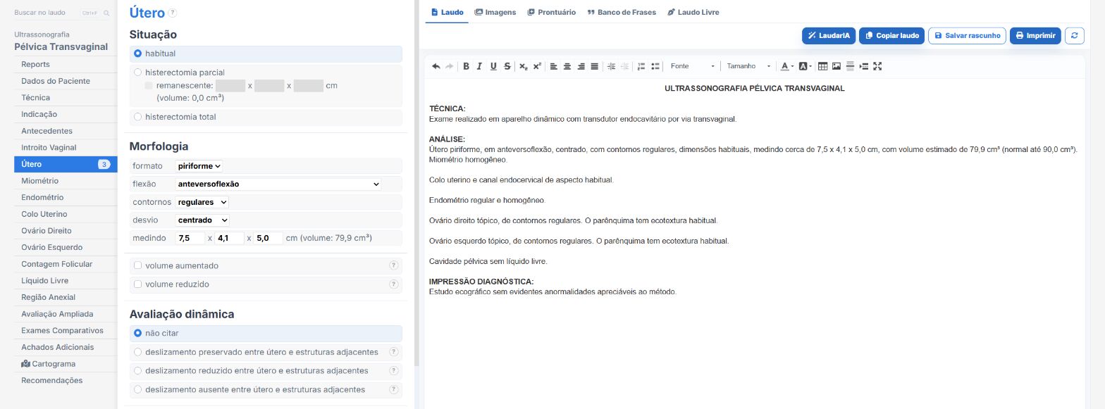
Laudário has templates for transvaginal and abdominal female pelvis, with and without Doppler, as well as endometriosis, saline infusion sonography, HyCoSy, ovarian reserve and follicular monitoring. In practice:
- Fibroids added one by one, with FIGO type and numbering.
- Adenomyosis by MUSA signs.
- Uterine and ovarian volume calculated.
- O-RADS classified from the descriptors.
- Recommendations in their own tab, separate from the findings.
Try it free for 15 days, no credit card required.
The ultrasound reporting platform built by sonologists. We write about reports, classifications and standardization from the real routine of the practice. Who writes and how.
Educational content for healthcare professionals. It does not replace clinical judgment or the guidelines adopted by your service. Editorial policy.