Thickened endometrium and endometrial polyp: what to measure, the right cutoffs and what to recommend
Endometrial thickness only makes sense with the cycle phase and menopausal status. The 4 mm cutoff and when it applies, the polyp's vascular pedicle, the differential with a submucosal fibroid and the red flags.
 LaudárioUltrasound reports in practice
This article is part of the guide Gynecologic ultrasound: the complete guide, from uterus to ovaries.
"Thickened endometrium" is one of the phrases that generates the most referrals for hysteroscopy, and also one of the most misinterpreted. Endometrial thickness only has meaning when you know which phase the patient is in: a 12 mm endometrium is expected in the secretory phase of a 30-year-old woman and is a red flag in a 60-year-old woman with bleeding.
This text shows how to measure, which values to use in each situation, how to describe the polyp and what to recommend.
How to measure
- Sagittal view of the uterus, at the point of greatest thickness.
- Measure both layers of the endometrium, from interface to interface with the myometrium.
- If there is fluid in the cavity, measure the two layers separately and add them, excluding the fluid.
- Record the measurement in millimeters, with the appearance (trilaminar, homogeneous, heterogeneous) and the regularity of the endometrial-myometrial interface.
An isolated measurement, without appearance and without context, is the main source of alarmist reports.
In the reproductive years: the cycle phase changes everything
In women who menstruate, the endometrium ranges from about 4 mm in the postmenstrual phase to 14 mm or more in the secretory phase. For this reason, the date of the last menstrual period should be in the report, and the trilaminar appearance (proliferative phase) or homogeneous hyperechoic appearance (secretory phase) helps with interpretation.
In the reproductive years there is no single thickness cutoff. What prompts investigation is a focal lesion (polyp, submucosal fibroid) or unexpected heterogeneity, and not the isolated number. When in doubt, repeating the exam right after menstruation, when the endometrium is thin, shows focal lesions better.
In postmenopause
Here the number matters, and the clinical context defines the cutoff:
- With bleeding: an endometrium of up to 4 mm, homogeneous and well visualized, has a high negative predictive value for endometrial cancer. Above that, or if poorly visualized, investigation (biopsy or hysteroscopy) is recommended.
- Without bleeding: the 4 mm cutoff does not apply directly, and slightly thicker endometria are frequent and usually benign. Higher values (many practices use around 11 mm) or focal findings justify investigation, depending on the context and risk factors.
- Hormone therapy and tamoxifen: they alter the endometrium. Tamoxifen produces a characteristic subendometrial cystic appearance, which should be described as such.
The report should state whether the patient is postmenopausal, whether there is bleeding and whether she uses hormones, because each combination has a different interpretation.
Endometrial polyp
It is the most common focal lesion of the cavity. The signs that identify it:
- Hyperechoic lesion, ovoid, well defined, within the cavity.
- Preserved endometrial interface around it.
- Single vascular pedicle on Doppler (the "feeding vessel"), the most characteristic sign.
- Sometimes, small cysts inside.
The report describes the location (wall, fundus, cornua), the measurements and the pedicle. Small polyps may not be seen against the thick endometrium of the secretory phase, and sonohysterography (with saline instillation into the cavity) greatly increases accuracy.
Polyp or submucosal fibroid?
- Polyp: hyperechoic, endometrium continuous over it, single vascular pedicle.
- Submucosal fibroid: hypoechoic, with shadowing, covered by endometrium, peripheral vascularization and origin in the myometrium.
The difference matters at hysteroscopy: the polyp is resected easily; a type 2 fibroid may require more than one surgical stage.
Red flags
Some findings suggest hyperplasia or neoplasia and should be highlighted in the diagnostic impression:
- Heterogeneous and irregular endometrium.
- Irregular or interrupted endometrial-myometrial interface.
- Diffuse vascularity or multiple vessels (multifocal) on Doppler.
- Collection in the cavity in a postmenopausal patient.
The IETA (International Endometrial Tumor Analysis) terminology standardizes the description of these findings and is worth using as a reference for the report.
Common errors
- "Thickened endometrium" without the cycle phase or menopausal status.
- Adding the fluid into the measurement.
- Polyp without the vascular pedicle.
- Applying the 4 mm cutoff to a patient without bleeding.
- Not recommending investigation when the endometrium is above the cutoff with bleeding.
See also our text on sonohysterography and HyCoSy.
How Laudário helps
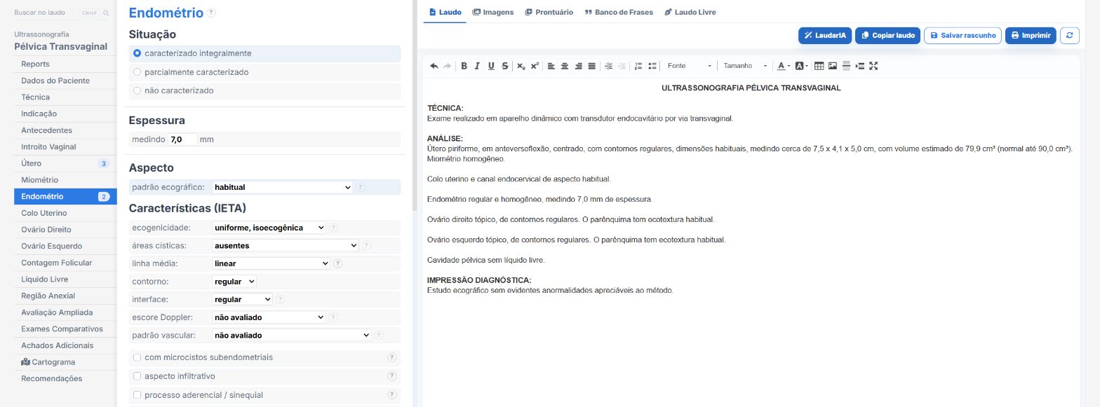
In the female pelvis templates of Laudário:
- Endometrial thickness and appearance with the cycle phase and menopausal status.
- Endometrial polyp with location, measurements and vascular pedicle.
- Sonohysterography template for evaluation of the cavity.
- Recommendations on their own tab.
Try it for 15 days free, no credit card required.
The ultrasound reporting platform built by sonologists. We write about reports, classifications and standardization from the real routine of the practice. Who writes and how.
Educational content for healthcare professionals. It does not replace clinical judgment or the guidelines adopted by your service. Editorial policy.