Infertility: sonohysterography and HyCoSy in the evaluation of the cavity and the tubes
Three questions, three exams, one window of the cycle that is not negotiable. See sonohysterography with Lasmar STEPW and FIGO, HyCoSy with the Cotte sign and periovarian spill, and antral follicle count, along with the pitfalls that make couples repeat the exam.
 LaudárioUltrasound reports in practice
This article is part of the guide Gynecologic ultrasound: the complete guide, from uterus to ovaries.
The infertility workup has three sonographic questions: is the uterine cavity normal? are the tubes patent? how much ovarian reserve remains? Three exams answer them (sonohysterography, HyCoSy and antral follicle count), and all three are done in the same office, with the same transducer, often in the same session.
What changes everything is timing: all three are only valid in a specific window of the cycle. Outside it, the exam is not imprecise, it is invalid. This text walks through all three, with the criteria, the classifications the surgeon expects and the pitfalls that make couples repeat the exam.
Timing: why the window is not negotiable
Sonohysterography and HyCoSy are performed in the early proliferative phase, typically between day 5 and day 10 of the cycle, right after menstrual flow ends. There are three reasons, and none of them is bureaucratic:
- The endometrium is thin. In the secretory phase it becomes thick and irregular, and easily mimics a polyp: a guaranteed false positive.
- There is no risk of pregnancy. Injecting contrast or saline into a cavity with an early pregnancy is the scenario the window exists to avoid.
- Flow has already stopped. Intracavitary blood and clots are the other classic source of false positives.
The contraindications are equally objective and must be checked beforehand: pregnancy (confirmed or possible), active pelvic infection or cervicitis, because the infusion can ascend the infection and cause PID, and significant active bleeding. Known hydrosalpinx deserves extra attention because of the risk of exacerbation.
Antral follicle count has its own, different window: the early follicular phase, between day 2 and day 5, when recruitment has not yet selected a dominant follicle and the count reflects the available pool.
Sonohysterography: the cavity under distension
Saline infusion sonohysterography (SIS) solves the central problem of the conventional transvaginal exam: the two walls of the cavity are touching each other. A small lesion is hidden between them. When saline separates the walls, each can be seen in isolation, and what was a nonspecific thickening becomes a polyp with a stalk, or nothing.
The technique: a thin Foley catheter (No. 8 is usually enough), balloon inflated with a small volume, infusion of 0.9% saline under real-time visualization, systematic sweep from the fundus to the isthmus in two planes. 3D adds the coronal plane, which is where malformations are revealed.
The degree of distension is a report finding
This is the equivalent of bowel preparation in endometriosis: a poorly distended cavity does not allow normality to be concluded. The report should record whether distension was adequate, partial (with segments of limited evaluation) or inadequate, and in the latter case, recommend repeat examination under better conditions instead of issuing a report that pretends to have seen.
Polyps
The findings that support the diagnosis: asymmetric focal thickening with the bright edge sign (the interface of the polyp with the endometrium), a single vascular stalk on Doppler (the most specific finding) and well-defined contours. Surrounded by fluid, the lesion appears as an endometrial nodule displacing the saline.
What the surgeon wants from the report, beyond the diagnosis, is the prediction of difficulty, and it comes from size and location:
- Less than 1 cm: low-complexity hysteroscopic polypectomy.
- 1 to 3 cm: intermediate complexity.
- Above 3 cm: high complexity; consider a resectoscope or morcellator.
A polyp with a well-defined stalk favors hemostasis; a location in the fundus or lateral walls makes access harder. These are sentences that fit in the report and change planning.
Submucosal fibroids: FIGO and Lasmar
Here the report needs to deliver two classifications, because they answer different questions.
FIGO describes the relationship with the myometrium:
- FIGO 0: entirely intracavitary, pedunculated.
- FIGO 1: submucosal with less than 50% intramural.
- FIGO 2: submucosal with 50% or more intramural.
Lasmar (STEPW) answers the question FIGO does not: can it be resected hysteroscopically? There are five components:
| Letter | What | 0 points | 1 point | 2 points |
|---|---|---|---|---|
| S | Size (largest diameter) | < 2 cm | 2 to 5 cm | > 5 cm |
| T | Extent of the base | ≤ 1/3 | 1/3 to 2/3 | > 2/3 |
| E | Location along the axis | lower third | middle third | upper third |
| P | Penetration into the myometrium | 0 (intracavitary) | < 50% | ≥ 50% |
| W | Wall | anterior/posterior | lateral | n/a |
The sum defines the group and the management:
- Score ≤ 4, Group I: low-complexity hysteroscopic myomectomy.
- Score 5 to 6, Group II: high complexity; consider pretreatment with a GnRH analogue and a two-step approach.
- Score ≥ 7, Group III: consider an alternative approach to hysteroscopy.
A report that delivers the calculated STEPW spares the hysteroscopist from redoing the math and, more importantly, avoids surgery started and interrupted for technical impossibility.
Synechiae, septa and what separates them
Synechiae appear as bands crossing the cavity, associated with incomplete and irregular distension: the saline cannot open the cavity because adhesions hold the walls together. It is worth classifying the type by site (endometrial, fibrotic or myometrial), which guides the difficulty of lysis.
In malformations, the finding that separates the entities is the external contour of the uterus, which is why the coronal plane of 3D is decisive:
- Arcuate: mild fundal indentation, less than 1 cm, angle greater than 90°, convex external contour. A normal variant.
- Septate: septum greater than 1.5 cm, angle less than 90°, convex external contour. It is correctable by hysteroscopy and is the malformation most associated with recurrent pregnancy loss.
- Bicornuate: serosal indentation greater than 1 cm, altered external contour. Not a case for hysteroscopic septoplasty.
Confusing septate with bicornuate leads to the wrong surgery. The external contour is the criterion.
It is worth mentioning the isthmocele (cesarean scar niche), increasingly common: record depth, length and, the finding that determines management, the residual myometrial thickness.
HyCoSy: tubal patency without radiation
Hysterosalpingo-contrast sonography answers the tubal question without ionizing radiation, without iodine and in the same office as the transvaginal exam. An echogenic contrast is used: the microbubble agent (sulfur hexafluoride) is the standard, and there is the alternative of a gel with saline and air.
The Cotte sign and the patency categories
The classic endpoint is the Cotte sign: positive when contrast passes through and spreads in the peritoneal cavity; negative when it does not. But a binary reading is insufficient, and the mature report works with more nuance:
- Patent: continuous progression from the ostium to the fimbria, with peritoneal spill. Cotte positive.
- Patent with slow progression: partial patency, reduced but present spill. Cotte positive, with a caveat.
- Proximal obstruction: contrast does not leave the ostial/interstitial segment. Cotte negative.
- Distal obstruction: contrast reaches the middle and distal segments but does not spill. Cotte negative.
- Tubal spasm: transient interruption. Cotte indeterminate.
The distinction between spasm and organic proximal obstruction is the classic problem of the exam, and it matters a great deal: spasm is functional and reversible; organic proximal obstruction may indicate tubal catheterization or in vitro fertilization. Spasm is most common precisely at the ostium, where the musculature is most reactive. When the exam is inconclusive, the report should say so and suggest a repeat study with analgesic and antispasmodic premedication, instead of asserting an obstruction that does not exist.
An error worth naming: unilateral tubal obstruction cannot be reliably diagnosed on a single exam. Preferential distension through the tube of lower resistance makes the other appear obstructed. A unilateral finding calls for caution in the wording.
Peritoneal spill and the periovarian pattern
Spill is the endpoint that confirms patency: free contrast in the pouch of Douglas and the paracolic gutters. And there is a fine sign worth looking for: the periovarian ring distribution, with contrast uniformly surrounding the ovary. A complete ring suggests not only patency but a preserved anatomical relationship between the fimbria and the ovary, which is, after all, what natural fertility requires. An incomplete arc or absence of distribution raises the possibility of peritubo-ovarian adhesions, even with a patent tube.
This is the point at which HyCoSy outperforms conventional hysterosalpingography: it sees the ovary, and it sees the relationship of the contrast to it.
Hydrosalpinx: the finding that changes treatment
Hydrosalpinx is not just a distal obstruction. It is a finding with its own strong therapeutic consequence: tubal fluid refluxing into the cavity reduces implantation rates in assisted reproduction, and the established management is salpingectomy or tubal occlusion before embryo transfer. A report that identifies hydrosalpinx and does not flag this implication delivered the finding without the information that justifies it.
Ovarian reserve: counting antral follicles
Antral follicle count (AFC) is the sonographic marker of reserve, counting follicles of 2 to 10 mm in both ovaries, in the early follicular phase. The ranges used in practice:
- 0 to 4: very low reserve; reduced response to stimulation expected.
- 5 to 8: low reserve.
- 9 to 19: normal reserve, with adequate response expected.
- Above 19: high reserve, with a possibly exaggerated response and increased risk of ovarian hyperstimulation syndrome.
Note that AFC is bidirectional: it serves not only to predict failure but also to predict an excess of response, and high reserve is the finding that leads the specialist to reduce the gonadotropin dose. It is safety information, not just prognosis.
AFC goes hand in hand with AMH, and the two complement each other. The report does not need to interpret the combination, but it must deliver the count per ovary and the total, because that is what goes into the dose calculation.
Pitfalls of infertility exams
- Performing the exam outside the proliferative phase: thick endometrium mimics a polyp, and the risk of pregnancy is no longer negligible.
- Not checking contraindications: active pelvic infection can ascend with the infusion.
- Not recording the degree of distension: a poorly distended cavity does not allow normality to be concluded.
- Diagnosing unilateral obstruction on a single exam: preferential distension misleads.
- Confusing spasm with proximal obstruction: the report can honestly be inconclusive and suggest a repeat study.
- Reporting hydrosalpinx without flagging the impact on implantation: the finding has its own management.
- Delivering a fibroid with FIGO only: FIGO describes; STEPW decides the route.
- Confusing septate with bicornuate: the external contour is the criterion, and the surgery is different.
- Counting antral follicles in the wrong phase: after recruitment, the count does not reflect the pool.
- Not measuring the residual myometrial thickness in isthmocele.
How Laudário helps with infertility reports
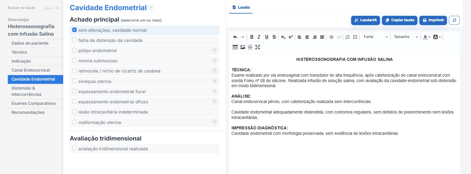
Laudário covers all three exams: Saline Infusion Sonohysterography, Hysterosalpingo-Contrast Sonography (HyCoSy), the Transvaginal + HyCoSy combination, as well as Ovarian Reserve and Follicular Monitoring. In practice:
- Lasmar STEPW classification calculated automatically from the fields, with the score, the group and the management sentence ready, and FIGO in parallel.
- Prediction of polypectomy complexity derived from size and location, written into the report.
- Mapping of synechiae by site, with an anatomical grid and automatic notation.
- Degree of distension as its own field, which alters the conclusion and flags a repeat exam when inadequate.
- In HyCoSy: patency in five categories (not binary), Cotte sign derived automatically from the patency chosen, peritoneal spill by site and periovarian distribution as ring/arc/absent.
- Contrast preparation with calculated volumes and an alert when the balloon falls outside the usual range.
- Consolidated bilateral conclusions ("bilateral tubal patency preserved" instead of two redundant sentences) and dedicated sentences for asymmetric combinations.
- Automatic recommendation about hydrosalpinx and implantation, and about repeat study with an antispasmodic when the finding is inconclusive.
- Follicle count per ovary and total, with automatic reserve classification and the hyperstimulation risk alert in high reserve.
- Support from LaudarIA for cases with multiple intracavitary findings.
If you handle infertility workups and put these reports together manually, it is worth trying: 15-day free trial, no credit card required, with all gynecologic and reproduction modules unlocked.
The ultrasound reporting platform built by sonologists. We write about reports, classifications and standardization from the real routine of the practice. Who writes and how.
Educational content for healthcare professionals. It does not replace clinical judgment or the guidelines adopted by your service. Editorial policy.