Polycystic ovaries on ultrasound: the updated criteria and how to report without causing alarm
Ovarian morphology is only one of the three PCOS criteria. The 20 follicles per ovary, the 10 mL volume, cycle phase, adolescents and how to write the finding without calling a follicle a cyst.
 LaudárioUltrasound reports in practice
This article is part of the guide Gynecologic ultrasound: the complete guide, from uterus to ovaries.
"Polycystic ovaries" is one of the phrases most often written in pelvic ultrasound reports, and one of those that causes the most confusion. To the patient, it sounds like a serious disease and "cysts." To the gynecologist, it only has value if it follows the right criterion, because ovarian morphology is just one of the three criteria for polycystic ovary syndrome (PCOS), and on its own it does not make the diagnosis.
This article shows how to count follicles, measure volume, apply the updated criteria and write the finding correctly and without alarm.
PCOS and where ultrasound fits in
By the Rotterdam criteria, PCOS is diagnosed with two of three findings, after excluding other causes:
- Oligo-ovulation or anovulation (irregular cycles).
- Clinical or biochemical hyperandrogenism.
- Polycystic ovarian morphology on ultrasound.
In other words, ultrasound delivers one criterion, not the diagnosis. For this reason, the report should describe the morphology and not conclude "polycystic ovary syndrome." The 2023 international guideline also reinforced that, in adolescents (within the first 8 years after menarche), ovarian morphology should not be used for diagnosis, because multifollicular ovaries are normal at this stage.
The morphology criteria
With high-frequency transducers (8 MHz or higher) by the transvaginal route, the current international guideline considers the following polycystic ovarian morphology:
- Follicle number per ovary of 20 or more (follicles of 2 to 9 mm), in at least one ovary; or
- Ovarian volume of 10 mL or more, in at least one ovary, in the absence of a cyst, corpus luteum or dominant follicle.
The old cutoff of 12 follicles was abandoned as equipment resolution improved, because normal ovaries began to exceed it. When the study is transabdominal, the count is unreliable and volume carries more weight.
How to measure
- Volume: three orthogonal diameters multiplied by 0.523 (ellipsoid formula). Record the volume of each ovary.
- Follicle count: sweep the entire ovary, counting all follicles of 2 to 9 mm. A common error is counting only those in a single plane.
- Cycle phase: ideally in the early follicular phase, between days 3 and 5. A dominant follicle (above 10 mm) or a corpus luteum increases the volume and invalidates the volume criterion for that ovary; in that case, the report should say so and the assessment can be repeated.
- Hormonal contraception: reduces volume and count, which should also be noted as a limitation.
Other findings that help
Some descriptive features are common in ovaries with polycystic morphology and can be recorded:
- Follicles distributed at the periphery, in a "string of pearls" pattern.
- Enlarged, hyperechoic central stroma.
They support the finding but do not replace the count and the volume, which are the objective criteria.
How to write without causing alarm
The word "cysts" for follicles is a frequent error and causes unnecessary anxiety. Follicles are normal structures. Appropriate wording:
- Describes the volume and follicle count of each ovary.
- Concludes with "ovaries with polycystic morphology" when the criteria are met, and not with the diagnosis of the syndrome.
- Recommends clinical and laboratory correlation, in the recommendations tab.
And when the count is high but the volume is normal, or the reverse, it is worth stating which criterion was met.
Antral follicle count and ovarian reserve
The same count has another use: the antral follicle count (AFC), summed across both ovaries, estimates ovarian reserve in patients undergoing fertility evaluation. A high AFC is typical of polycystic morphology and is associated with a risk of hyperstimulation syndrome in assisted reproduction treatments, useful information for the specialist.
Common errors
- Concluding "polycystic ovary syndrome" from ultrasound.
- Using the 12-follicle cutoff.
- Measuring volume with a dominant follicle or corpus luteum present.
- Calling follicles cysts.
- Applying the criterion in adolescents.
See also our article on the transvaginal pelvic report.
How Laudário helps
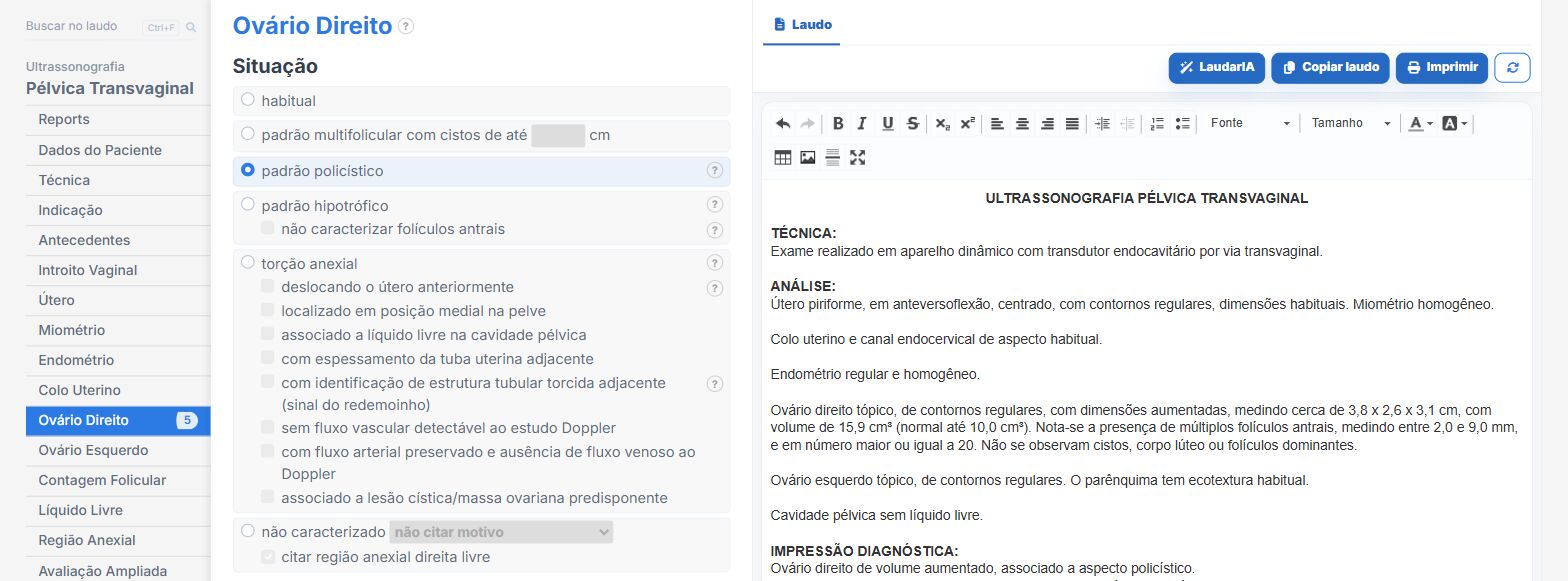
In the female pelvis and ovarian reserve templates of Laudário:
- Ovarian volume calculated automatically from the three diameters.
- Follicle count per ovary and antral follicle count.
- Polycystic morphology described by the criteria, without turning the finding into a diagnosis.
- Recommendations in their own tab.
Try it free for 15 days, with no credit card.
The ultrasound reporting platform built by sonologists. We write about reports, classifications and standardization from the real routine of the practice. Who writes and how.
Educational content for healthcare professionals. It does not replace clinical judgment or the guidelines adopted by your service. Editorial policy.