Follicular monitoring on ultrasound: what to measure and how to record progress
How to plan the serial studies of follicular monitoring, how to measure the dominant follicle and the endometrium, which signs indicate ovulation and how to organize the report so the specialist can follow the cycle at a glance.
 LaudárioUltrasound reports in practice
This article is part of the guide Gynecologic ultrasound: the complete guide, from uterus to ovaries.
Follicular monitoring follows the growth of the follicles and endometrium throughout the cycle with serial ultrasounds. It is used in natural cycles, timed intercourse, intrauterine insemination and ovarian stimulation. The specialist decides the timing of ovulation and management from these numbers, so the measurement must always be done the same way and the report must show the progression.
The baseline study
At the start of the cycle, usually between day 2 and day 4, the baseline study confirms there are no functional cysts that would interfere with stimulation, records the thin endometrium of the menstrual phase and, when requested, the antral follicle count.
The serial studies
- They begin around day 8 to 10, or according to the specialist's protocol.
- The dominant follicle usually stands out from about 10 mm and grows, on average, close to 1.5 to 2 mm per day.
- The preovulatory follicle, in a natural cycle, is usually around 18 to 24 mm. With medication, the reference values change and the specialist defines the timing of the trigger.
How to measure
- Each follicle is measured in two or three orthogonal diameters, and the report gives the mean diameter.
- Follicles are listed by ovary, from largest to smallest. Small follicles can simply be counted.
- The endometrium is measured in the longitudinal view, at its greatest thickness, and described by pattern: the trilaminar pattern usually accompanies the late follicular phase.
Signs of ovulation
- Disappearance or reduction of the dominant follicle.
- Formation of a corpus luteum, with thick walls and peripheral vascularity on Doppler.
- A small amount of free fluid in the cul-de-sac.
Situations that should be highlighted
- Many large follicles in a low-complexity cycle, because of the risk of multiple pregnancy and hyperstimulation.
- A follicle that grows and does not rupture (luteinized unruptured follicle).
- A cyst that persists since the baseline study.
- Thin endometrium or one with a focal lesion (polyp).
The report: one table per study
What helps the specialist most is seeing the whole cycle at a glance. Each study should include the date, the cycle day, the medication in use when reported, the follicles of each ovary with the mean diameter, and the endometrial thickness and pattern. In table format, the progression appears without having to open the previous reports.
Common errors
- Measuring the follicle in a single diameter.
- Not reporting the cycle day.
- Mixing follicles from both ovaries in a single list.
- Failing to mention the free fluid and the corpus luteum after ovulation.
- Not highlighting an excess of large follicles.
See also our article on saline infusion sonography and HyCoSy in infertility.
How Laudário helps
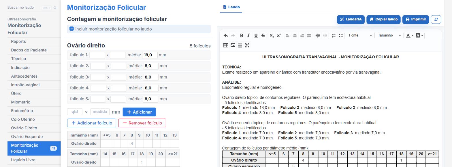
In the follicular monitoring template of Laudário:
- Ovaries, follicles and endometrium in their own tabs, within the transvaginal report.
- Measurements in their own fields, with the sentence built from them.
- Recommendations in their own tab.
Try it free for 15 days, no credit card required.
The ultrasound reporting platform built by sonologists. We write about reports, classifications and standardization from the real routine of the practice. Who writes and how.
Educational content for healthcare professionals. It does not replace clinical judgment or the guidelines adopted by your service. Editorial policy.