Transvaginal Pelvic Ultrasound: How to Structure the Gynecologic Report from Uterus to Adnexa
How to structure the transvaginal pelvic ultrasound report: uterus, endometrium, ovaries, IOTA/O-RADS classifications and the most common mistakes that compromise management.
 LaudárioUltrasound reports in practice
This article is part of the guide Gynecologic ultrasound: the complete guide, from uterus to ovaries.
Transvaginal pelvic ultrasound is one of the most frequent exams in gynecologic practice and, at the same time, one of those that suffers most from generic reports. Small omissions, such as an endometrium described without the cycle phase, an ovary without a volume, or an adnexal mass without adequate morphology, turn a potentially decisive exam into a document of little use to the gynecologist. In this article, we propose a practical structure for the transvaginal pelvic ultrasound report, covering the uterus, endometrium, ovaries and the main classifications in use (IOTA, O-RADS, FIGO for fibroids), as well as the most frequent mistakes that undermine the clinical usefulness of the exam.
When transvaginal pelvic ultrasound is indicated
Transvaginal ultrasound is the route of choice for gynecologic evaluation in sexually active patients, because it offers far better resolution than the abdominal route for the uterus, endometrium and adnexa. The main indications in outpatient practice include:
- Abnormal uterine bleeding (AUB): investigation of polyps, submucosal fibroids, endometrial hyperplasia and endometrial neoplasia, especially in postmenopause.
- Acute or chronic pelvic pain: ovarian cysts, pelvic inflammatory disease, deep endometriosis, adnexal torsion and adenomyosis.
- Infertility workup: evaluation of uterine morphology, antral follicle count, signs of PCOS and indirect tubal patency.
- Evaluation of pelvic masses: morphologic and vascular characterization of adnexal lesions, applying the IOTA and O-RADS criteria.
- IUD follow-up: checking position, mainly after insertion or in patients with symptoms attributed to the device.
- Pre- and postoperative gynecologic care: planning of myomectomy, hysteroscopy and endometriosis resection, and follow-up after the procedure.
Each of these indications calls for different emphasis in the report. An exam for AUB needs to characterize the endometrium in the greatest possible detail and correlate it with the cycle phase. An infertility workup needs a precise antral follicle count and a careful description of the uterus. When the report does not speak to the clinical indication, the gynecologist loses time and the patient ends up repeating exams or delaying management.
Technical aspects before typing the report
Before describing findings, it is important to record in the technique block some information that completely changes the interpretation:
- Menstrual cycle phase or menopausal status: essential for correct reading of the endometrium and ovaries.
- Hormone use: combined oral contraceptives, progestins, replacement therapy, tamoxifen or GnRH agonists radically change the endometrial and ovarian appearance.
- Bladder emptying: the transvaginal exam requires an empty bladder; a full bladder distorts the anatomy and hinders the scan.
- Technical limitations: patients who have never been sexually active (in whom a transrectal or suprapubic-only approach is chosen), marked retroversion, surgical scars or intolerance to the exam should be recorded to give context to what could be evaluated.
- Comparison with prior exams: when available, citing the uterine volume, endometrial thickness and ovarian dimensions of the previous exam makes follow-up easier.
Recommended structure for the report
1. Uterus: morphology, position and measurements
Describe the position (anteverted-anteflexed, midposition, retroverted-retroflexed), the contours (regular or bulging because of fibroids), the myometrial echotexture (homogeneous or heterogeneous, with or without areas suggestive of adenomyosis) and the three measurements (longitudinal, anteroposterior and transverse). State the estimated volume in cubic centimeters, ideally by the ellipsoid formula. In postmenopausal patients, record whether the uterus is atrophic as expected for the age group.
2. Myometrium and adenomyosis
When adenomyosis is suspected, use the MUSA criteria (Morphological Uterus Sonographic Assessment): myometrial asymmetry, myometrial cysts, hyperechoic islands, fan-shaped shadowing, translesional vascularity, and an interrupted or irregular junctional zone. Describe what is present instead of using only the term “suggestive of adenomyosis”: a structured description lets the gynecologist weigh the finding against the clinical picture.
3. Fibroids: location and FIGO classification
For each relevant fibroid, report the location (anterior, posterior, fundal, lateral or isthmic wall), dimensions in three planes, echogenicity, presence of degeneration and, above all, the FIGO 0-8 classification. The FIGO classification is what guides surgical management: FIGO 0 (pedunculated intracavitary) and FIGO 1 (submucosal with less than 50% intramural component) fibroids are usually resectable by hysteroscopy; FIGO 2 to 5 fibroids often require a laparoscopic or open approach. Citing only “presence of fibroids” without classifying them drastically reduces the usefulness of the report.
4. Endometrium
Measure the endometrial thickness in a sagittal plane, from the outer edge of one basal layer to the outer edge of the opposite one, at the greatest visible thickness. Describe the echotexture (trilaminar, homogeneous hyperechoic, heterogeneous), the regularity of the endometrial-myometrial interface, the presence of fluid in the cavity and of focal images (polyps, submucosal fibroids). Always relate the thickness to the cycle phase or menopausal status: in postmenopause, values above 4-5 mm in patients with bleeding require investigation. In patients taking tamoxifen, the appearance may be cystic and exuberant even in the absence of pathology.
5. Ovaries: morphology, volume and follicles
Report the location (in the ovarian fossa, displaced, not identified), the three measurements and the volume calculated by the ellipsoid formula. Describe the presence of follicles, an approximate count when investigating PCOS or ovarian reserve, and the appearance of the stroma. In women of reproductive age, the antral follicle count (AFC) is an increasingly valued measure: report it when requested and state whether the count was done by three-dimensional or two-dimensional scanning.
6. Adnexal masses: applying IOTA and O-RADS
Adnexal masses deserve a detailed description: dimensions, content (anechoic, with septations, with a solid component), septation thickness, presence of papillary projections, color Doppler vascularity and vascular distribution. The IOTA Simple Rules and O-RADS US classifications structure this description into risk categories (from O-RADS 1, physiologic cyst, to O-RADS 5, high probability of malignancy), standardizing management. A report that merely describes a “complex adnexal cyst” without applying formal criteria misses the chance to directly guide the gynecologist on the urgency of referral.
7. Pouch of Douglas, sliding sign and deep endometriosis
In patients with chronic pelvic pain or suspected endometriosis, assess free fluid in the pouch of Douglas, the sliding sign between the rectosigmoid and the posterior uterine wall (a negative sign suggests adhesions) and the presence of hypoechoic nodules in the uterosacral ligaments, parametria, rectovaginal septum or bowel wall. An exam with bowel preparation considerably increases accuracy for deep endometriosis, so state in the report whether this preparation was performed.
8. IUD assessment, when applicable
In patients with an IUD, describe the type (copper or hormonal), the position (uterine body, displaced to the isthmus or cervix, with or without perforation) and the distance from the upper end to the fundus of the endometrial cavity. Displaced IUDs explain a large share of bleeding or pain complaints.
9. Diagnostic impression
The conclusion should restate the main findings in direct language and correlate them with the indication. In AUB, mention endometrial thickness, focal findings and the FIGO classification of fibroids. In an infertility workup, report uterine morphology, AFC and signs of PCOS. In adnexal masses, finish with the O-RADS category. The more the conclusion speaks to the reason for the exam, the more value the report delivers.
Common mistakes in the transvaginal pelvic ultrasound report
Even among experienced professionals, a few oversights frequently compromise the quality of the gynecologic report:
- Not stating the cycle phase: without this information, reading the endometrium and ovaries is impaired and the gynecologist has to infer.
- Fibroids without FIGO classification: surgical management depends on this classification; describing only a generic location is not enough.
- Endometrium measured in an oblique plane: measurements outside a perfect sagittal plane can overestimate the thickness and lead to unnecessary additional tests.
- Adnexal masses without IOTA/O-RADS: the absence of a risk category leaves the gynecologist to reconstruct the criteria, a task that belongs to the person performing the exam.
- Adenomyosis described generically: using “suggestive of adenomyosis” without citing which MUSA criteria are present empties the finding of meaning.
- Generic conclusion: “exam within normal limits” in a patient with chronic pelvic pain, without commenting on the pouch of Douglas, sliding sign and uterosacral ligaments, does not answer the referring physician's question.
- Poor comparisons: citing a prior exam without restating uterine dimensions, endometrial thickness and ovarian volumes does not allow longitudinal follow-up.
How Laudário helps structure transvaginal pelvic reports
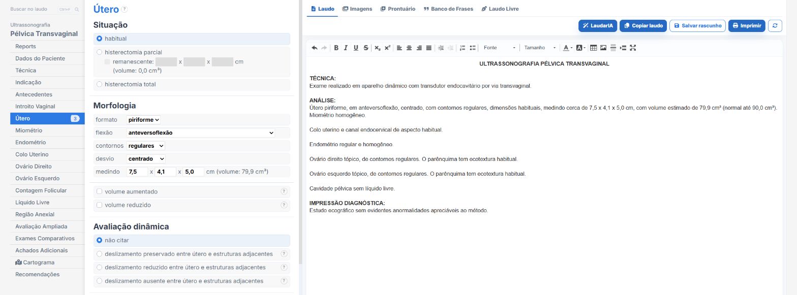
Laudário was designed for professionals who face this routine every day. For transvaginal pelvic ultrasound, and for the main gynecologic protocols, the system offers:
- Ready-made, structured templates for transvaginal gynecologic, suprapubic pelvic, infertility, PCOS, endometriosis with bowel preparation and adnexal mass follow-up, organized in the natural scanning order.
- Standardized, customizable phrases for uterus, endometrium, fibroids, ovaries, pouch of Douglas and sliding sign, keeping terminology consistent across reports of the same patient.
- Automatic calculations integrated into the typing workflow, including uterine volume, ovarian volume, endometrial thickness, antral follicle count and O-RADS categorization, avoiding manual math and transcription errors.
- Classifications already built in to the templates, with FIGO 0-8 for fibroids, MUSA for adenomyosis and IOTA/O-RADS for adnexal masses: you just select the scenario and the system delivers the matching phrase.
- A complete library covering pelvic, obstetric (1st, 2nd and 3rd trimester), abdominal, thyroid, breast, prostate and Doppler exams, all in a single online system.
The result is simple: less time typing, fewer forgotten findings and more consistent reports across exams of the same patient, which makes all the difference in following women under investigation for AUB, infertility, endometriosis or adnexal mass surveillance. Instead of starting from a blank page with each patient, you adjust a template that has already been tested, keep the clinic's professional standard and focus on what really demands your attention: reading the images.
If you still build your pelvic reports manually or by copying old text, it is worth getting to know Laudário. Try it free for 15 days and see in practice how a well-designed structure saves time on every exam.
The ultrasound reporting platform built by sonologists. We write about reports, classifications and standardization from the real routine of the practice. Who writes and how.
Educational content for healthcare professionals. It does not replace clinical judgment or the guidelines adopted by your service. Editorial policy.