O-RADS on ultrasound: how to classify adnexal masses and stop reporting "complex cyst"
ACR O-RADS replaces "complex cyst" with an objective lexicon and five categories with defined risk and management. See the descriptors, the 1 to 4 color score, the 10 cm cutoff, the typical lesions and what changes after menopause.
 LaudárioUltrasound reports in practice
This article is part of the guide Standardized classifications in the report: a guide to BI-RADS, TI-RADS and O-RADS.
“Complex cyst” is probably the most dangerous term in the gynecologic report. It is not a diagnosis, not a category, carries no estimated risk and does not indicate management. It merely tells the requesting physician that the sonologist saw something that was not simple, and it hands the whole decision to someone who did not see the image. The result is predictable: some of these patients are operated on unnecessarily, and others are followed when they should have been referred.
ACR O-RADS US (Ovarian-Adnexal Reporting and Data System) exists to end this. Like BI-RADS in the breast and TI-RADS in the thyroid, it offers a standardized lexicon of descriptors and a ladder of categories from 0 to 5, each with an estimated risk of malignancy and an associated management. The promise is simple and strong: if you describe the lesion with the right descriptors, the category comes out on its own, and with it, the management.
Why the lexicon solves the problem
The central point of O-RADS is not the category table. It is the lexicon. The reason “complex cyst” survives in reports is that it is a shortcut for avoiding a choice between descriptors. Once the examiner is required to answer the lexicon questions one by one, the category stops being an opinion and becomes a consequence.
The questions are these, in this order:
- Is the lesion cystic, solid, or almost solid (≥ 80% solid component)?
- Is there a solid component? Absent or present.
- How many locules? Unilocular, bilocular or multilocular.
- Are the margins (of the lesion or of the solid component) smooth or irregular?
- If there is a solid component, is it papillary? Fewer than 4 papillary projections, 4 or more projections, or a nonpapillary solid component.
- What is the Doppler color score? From 1 to 4.
- What is the largest diameter?
Note that none of these questions accepts “complex” as an answer. It is a lexicon designed to leave no escape route.
The complementary descriptors
Besides the structural ones, some descriptors help recognize typical lesions and should appear in the description when present:
- Posterior acoustic shadowing: suggests a dense component, characteristic of mature teratoma and some fibromas.
- Low-level internal echoes: the ground-glass appearance of the endometrioma.
- Incomplete septations: septa that do not completely cross the lesion, a typical finding of hydrosalpinx and peritoneal inclusion cyst.
- Reticular pattern or retracting clot: the signature of the hemorrhagic cyst.
- Rokitansky nodule, echogenic lines and dots, fat-fluid level: the constellation of the dermoid.
The color score: a number, not an adjective
The color score is often mishandled in reports, reduced to “vascularized” or “poorly vascularized.” In O-RADS it is an ordinal scale from 1 to 4, assessed in the solid component or in the most vascularized wall/septum:
- Color score 1: avascular, no detectable flow.
- Color score 2: scant vascularity (minimal flow).
- Color score 3: moderate vascularity.
- Color score 4: marked vascularity.
There is an operational subtlety worth internalizing: the color score does not always decide anything. In a unilocular cystic lesion with no solid component, the score is 1 by definition, since there is nothing to vascularize. In a solid lesion, the score is what separates category 3 from 4 and 5. Knowing when the score is decisive and when it is merely descriptive avoids both excess and omission.
The categories: risk and management
O-RADS 0: incomplete evaluation
A rarely used and useful category. It applies when the study is technically insufficient to characterize the lesion: the patient did not tolerate the transvaginal approach, the window was inadequate, or the lesion was not completely visualized. Formally recognizing this is better than misclassifying. The management is to repeat the study under adequate conditions or to complement it with another method.
O-RADS 1: normal ovary (virtually no risk)
This is not a lesion: it is the physiologic ovary of the premenopausal patient. Here belong the follicle (up to 3 cm), the corpus luteum and the corpus albicans. None of them is a lesion, none of them needs to be classified as 2 to 5, and none of them justifies follow-up.
A highlight on the corpus luteum, which is responsible for a disproportionate number of unnecessary referrals: thick, regular walls, with the characteristic peripheral vascularity in a ring of fire pattern on Doppler. An examiner who does not recognize the ring of fire may read a hemorrhagic corpus luteum as a vascularized lesion with a worrying appearance, and the patient ends up in an MRI. The most useful practical rule here is temporal: the corpus luteum is physiologic and transient, and a repeat study in another phase of the cycle resolves the doubt at no cost.
Remember that, in postmenopause, follicles and corpora lutea do not exist. An image with these characteristics in a postmenopausal patient is not physiologic and needs to be reclassified.
O-RADS 2: almost certainly benign (risk < 1%)
This is the category of benign lesions with typical appearance, and the size ceiling is 10 cm. It includes:
- Simple cyst: unilocular, anechoic, thin smooth walls, no solid component, avascular.
- Hemorrhagic cyst: unilocular, with internal echoes in a reticular pattern or with a retracting clot, avascular.
- Endometrioma: unilocular, smooth walls, fine low-level echoes (ground glass), no solid component, avascular.
- Mature teratoma (dermoid): echogenic component with acoustic shadowing, Rokitansky nodule, echogenic lines and dots, fat-fluid level, no significant vascularity.
- Paraovarian cyst: extraovarian, unilocular, anechoic, with the ovary identified separately.
- Peritoneal inclusion cyst: a collection that molds to the pelvic structures, surrounding the ovary without displacing it.
- Hydrosalpinx: anechoic tubular structure with incomplete septations.
- Unilocular or bilocular cystic lesion with smooth walls and no solid component, smaller than 10 cm.
Management in premenopause is, in general, none: these are benign findings that do not need follow-up. In postmenopause, the threshold is more conservative: ultrasound surveillance according to type and size, at clinical discretion.
Note the underlying logic: when a typical lesion exceeds 10 cm, it moves up to O-RADS 3. Not because its nature has changed, but because size reduces confidence in the typification and carries its own implications (risk of torsion, compressive symptoms, difficulty with complete characterization).
O-RADS 3: low risk (approximately 1% to 10%)
This is the category of the low-risk indeterminate. It includes:
- Unilocular cystic lesion with irregular margins, no solid component.
- Unilocular or bilocular cystic lesion with smooth walls measuring 10 cm or more.
- Multilocular cystic lesion with smooth margins, no solid component, smaller than 10 cm and color score 1 to 3.
- Solid lesion with smooth margins and color score 1.
- Any typical lesion (endometrioma, dermoid, hemorrhagic) above 10 cm.
Management: ultrasound surveillance or complementation with magnetic resonance imaging, considering evaluation by a sonologist with expertise in adnexal masses. In postmenopause, the threshold for complementing and referring is lower.
A note on the solid lesion with color score 1: it falls in O-RADS 3, which sometimes surprises. This is the typical category of the ovarian fibroma: solid, smooth margins, with acoustic shadowing and practically avascular. It is solid, but it is benign, and the color score is what supports this reading.
O-RADS 4: intermediate risk (approximately 10% to 50%)
Here the lesion starts to require specialized evaluation. It includes:
- Unilocular cystic lesion with a solid component and fewer than 4 papillary projections.
- Unilocular cystic lesion with a nonpapillary solid component and color score 1 or 2.
- Multilocular cystic lesion without a solid component measuring 10 cm or more, or with color score 4.
- Multilocular cystic lesion with irregular margins without a solid component.
- Bilocular or multilocular cystic lesion with a solid component and color score 1 or 2.
- Solid lesion with smooth margins and color score 2 or 3.
Management: magnetic resonance imaging and/or specialized gynecologic evaluation, considering referral to gynecologic oncology.
O-RADS 5: high risk (approximately 50% or more)
- Unilocular cystic lesion with a solid component and 4 or more papillary projections.
- Unilocular cystic lesion with a nonpapillary solid component and color score 3 or 4.
- Bilocular or multilocular cystic lesion with a solid component and color score 3 or 4.
- Solid lesion with irregular margins, regardless of color score.
- Solid lesion with smooth margins and color score 4.
- Presence of ascites and/or peritoneal implants: this is the criterion that overrides all the others.
Management: evaluation in gynecologic oncology; MRI may help with treatment planning.
Two shortcuts deserve emphasis because they simplify the decision in practice. The first: in a solid lesion, irregular margins lead straight to 5; the color score does not rescue it. The second: ascites and peritoneal implants are O-RADS 5 by themselves, regardless of how the adnexal lesion is characterized. No descriptor of the mass undoes these two findings.
Premenopause and postmenopause: what changes and what does not
There is a frequent misunderstanding here. Menopausal status does not change the classification criteria: the decision tree is the same, the descriptors are the same, the size cutoffs are the same. What changes is the clinical reading around it:
- Physiologic findings cease to exist. Follicles and corpora lutea are incompatible with postmenopause; the same image that would be O-RADS 1 at age 30 needs to be classified at age 60.
- The management threshold is lower. An O-RADS 2 that would not need follow-up in premenopause deserves surveillance according to type and size. An O-RADS 3 has a lower threshold for complementation and specialized evaluation.
- The reference ovarian volume falls. Approximately 10 cm³ in the reproductive years versus about 5 cm³ in postmenopause, which changes what is considered an enlarged ovary.
- The pretest probability rises. The same category carries greater clinical concern, even though the tabulated risk is the same.
It is a defensible design: the category describes the image; the context modulates the management. But it requires the report to state the menopausal status; without it, the recommendation is left hanging.
What to do with the cystadenoma
A paragraph is worth devoting to the lesion that most often becomes a “complex cyst”: the cystadenoma. The serous cystadenoma is usually unilocular, thin-walled and anechoic, and falls comfortably in O-RADS 2 when smaller than 10 cm. The mucinous one is typically multilocular, sometimes bulky, with content of variable echogenicity between the locules, and falls in O-RADS 3 or 4 depending on size, color score and presence of a solid component.
The point is that neither needs the word “complex”. The lexicon covers it: number of locules, presence of a solid component, margins, color score, size. Described this way, the bulky mucinous cystadenoma comes out as O-RADS 3 with a recommendation for complementation, which is exactly the correct management and exactly what “complex cyst” does not convey.
Pitfalls in the adnexal mass report
- Writing “complex cyst”: the term has no associated risk, no management and hands the decision to someone who did not see the image.
- Classifying the corpus luteum: it is physiologic; the ring of fire is the signature, and repeating the study in another phase of the cycle resolves the doubt.
- Omitting the largest diameter: without the measurement, the category cannot be assigned. It is better not to classify than to guess.
- Reporting “vascularized” instead of the color score: the 1 to 4 score is a classification criterion, not an adjective.
- Not counting the papillary projections: fewer than 4 is O-RADS 4; 4 or more is O-RADS 5. It is a count, not an impression.
- Ignoring ascites and implants: they are O-RADS 5 by themselves and override any descriptor of the mass.
- Failing to classify per lesion: when there is more than one, each lesion has its own category, and the management must address all the categories present, not just the largest.
- Describing an endometrioma as a “cyst with thick content”: it has a typical description and its own category.
- Not stating the menopausal status: without it, the recommendation cannot be calibrated.
- Classifying without citing the descriptors that support the category: a report that says “O-RADS 4” without describing why allows neither audit nor second opinion.
How Laudário helps classify adnexal masses
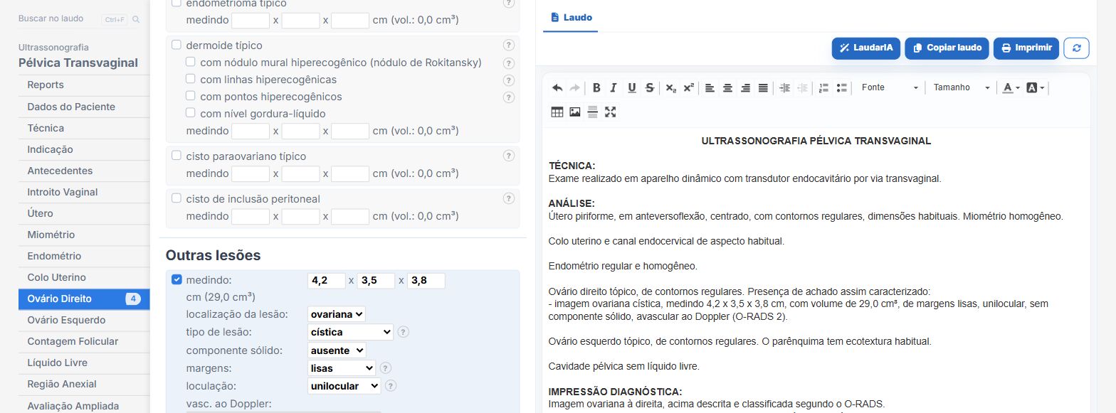
Laudário integrates O-RADS into the ovary module of the transvaginal pelvic report, with the classification derived automatically from the descriptors rather than chosen by hand. In practice, this means:
- The complete lexicon in dedicated fields: lesion type, solid component, loculation, margins, papillary projections, color score, acoustic shadowing, low-level echoes and incomplete septations. No room for “complex.”
- O-RADS category calculated automatically from the combination of descriptors and the largest diameter, following the ACR decision tree, including the shortcuts of irregular margins in a solid lesion and the 10 cm cutoff.
- Fields that appear only when they are decisive: in a unilocular cystic lesion with no solid component, the color score is fixed at 1 automatically; in a multilocular lesion with a solid component, the papillae field is hidden because it does not change the result. Fewer clicks, less noise, fewer errors.
- Typical lesions with ready-made phrases and automatic category (simple cyst, hemorrhagic cyst (reticular or retracting clot), endometrioma, dermoid with all its descriptors, paraovarian cyst, peritoneal inclusion cyst and hydrosalpinx), all with the 10 cm cutoff applied automatically.
- Physiologic findings handled as such: follicle, corpus luteum (with the ring of fire described) and corpus albicans receive no category, and are automatically blocked when the patient is registered as postmenopausal.
- Recommendations issued per category present, with text adjusted to menopausal status, including the lower threshold for complementation in postmenopause.
- A dynamic lesion adder, with independent classification per lesion and per ovary, and automatic aggregation of the categories in the recommendations block.
- No measurement, no category: the system does not assign O-RADS when the diameter has not been entered, rather than risking a guess.
- Support from LaudarIA, the integrated artificial intelligence, to help write difficult cases: bulky multilocular lesions, bilateral findings or patients in follow-up.
The result is a report that the gynecologist can use immediately: the lesion described with the right descriptors, the category derived from them and the associated management, all in the same document. In an area where the decision swings between doing nothing and referring to oncology, this explicit chain from descriptor to category to management is what protects the patient from both ends of the error.
If you still write “complex cyst” in your reports (and most services still do), it is worth trying: Laudário offers a 15-day free trial, with no credit card, with immediate access to the transvaginal pelvic report with integrated O-RADS, the endometriosis module and all the other protocols of the gynecologic routine.
The ultrasound reporting platform built by sonologists. We write about reports, classifications and standardization from the real routine of the practice. Who writes and how.
Educational content for healthcare professionals. It does not replace clinical judgment or the guidelines adopted by your service. Editorial policy.