Thyroid Ultrasound and TI-RADS: How to Describe and Classify Nodules in the Report
ACR TI-RADS scores five morphologic categories of the thyroid nodule and assigns a TR1 to TR5 level, which, combined with lesion size, indicates FNA, follow-up or no action. See how to structure the report.
 LaudárioUltrasound reports in practice
This article is part of the guide Standardized classifications in the report: a guide to BI-RADS, TI-RADS and O-RADS.
In thyroid ultrasound, TI-RADS (Thyroid Imaging Reporting and Data System) is the system that turns the nodule description into an objective management recommendation. In the most widely used version today, the ACR TI-RADS (American College of Radiology, 2017), each nodule receives points in five morphologic categories (composition, echogenicity, shape, margin and echogenic foci), and the sum defines the final category, from TR1 to TR5. That category, combined with lesion size, indicates whether the case calls for FNA (fine-needle aspiration), ultrasound follow-up or neither. In short: TR1 and TR2 require neither FNA nor follow-up; TR3 calls for FNA from 2.5 cm and follow-up from 1.5 cm; TR4, FNA from 1.5 cm and follow-up from 1.0 cm; TR5, FNA from 1.0 cm and follow-up from 0.5 cm.
For this classification to work, the report must describe each relevant nodule with location, dimensions in all three axes and the five scored features, state the total score, assign the TR category and close the conclusion by explicitly saying whether or not FNA is indicated. With nodules present in about half of adults undergoing ultrasound, the challenge is no longer detecting the lesion but stratifying risk and standardizing the recommendation, avoiding unnecessary punctures in very low-risk lesions without missing the patient who truly needs workup. Below, the complete report structure, category by category.
Why the thyroid report demands so much standardization
The thyroid is a small, superficial gland that is relatively simple to scan, but the clinical impact of the report is large, because every nodule described will or will not generate an FNA indication. A nodule described as "hypoechoic, solid, with microcalcifications" without a risk category leaves doubt about management. A nodule simply called "suspicious" without morphologic detail cannot be compared with future exams. And a conclusion that recommends FNA without objective criteria exposes the patient to potentially avoidable invasive procedures.
TI-RADS standardization solves three problems at once: it standardizes the morphologic description, stratifies the risk of malignancy and guides the FNA indication based on lesion size. It is the same reasoning as BI-RADS in the breast: turning a historically subjective assessment into an objective, auditable language understood both by the person reading the images and by the endocrinologist who receives the document. For the sonologist, this means a report that is more technically defensible and clinically more useful. For the patient, fewer repeated exams and fewer unnecessary procedures.
Technique and limitations: the block that opens the report
Before the anatomic description, it is worth recording the type of exam, the equipment when relevant and any technical limitation observed: patient body habitus, a gland that is difficult to access, a diving (substernal) goiter or prior surgery. This block gives context to the referring physician and protects the professional who signs.
The standard description of the thyroid parenchyma
Before getting to the nodules, the report must characterize the gland as a whole:
- Topography and dimensions: orthotopic or ectopic thyroid, with measurements of the right and left lobes (longitudinal, transverse and anteroposterior) and of the isthmus. Gland volume can be calculated with the ellipsoid formula (length x width x thickness x 0.524) for each lobe.
- Contours, echotexture and echogenicity: homogeneous or heterogeneous, normoechoic, hypoechoic or hyperechoic relative to the adjacent neck musculature. Diffuse heterogeneity associated with hypoechogenicity suggests chronic thyroiditis and should be stated explicitly.
- Vascularity on color Doppler: normal, increased or reduced. Useful mainly in the setting of thyroiditis and diffuse goiters.
- Cervical lymph nodes: in cases with suspicious nodules, scanning of the cervical chains (levels II to VI) should be systematic, with description of morphology, hilum, vascularity and measurements of atypical nodes.
The five morphologic categories of ACR TI-RADS
Knowing the five categories is what ensures a consistent report. The description of each nodule should follow this same sequence, so that the final category is a natural consequence of the text.
| Category | Finding | Points |
|---|---|---|
| Composition | cystic or almost completely cystic | 0 |
| spongiform | 0 | |
| mixed cystic and solid | 1 | |
| solid or almost completely solid | 2 | |
| Echogenicity | anechoic | 0 |
| hyperechoic / isoechoic | 1 | |
| hypoechoic | 2 | |
| very hypoechoic | 3 | |
| Shape | wider than tall | 0 |
| taller than wide | 3 | |
| Margin | smooth or ill-defined | 0 |
| lobulated or irregular | 2 | |
| extrathyroidal extension | 3 | |
| Echogenic foci | none or large comet-tail artifacts | 0 |
| macrocalcifications | 1 | |
| peripheral (rim) calcifications | 2 | |
| punctate echogenic foci (microcalcifications) | 3 |
A few points deserve attention when scoring. Spongiform nodules, formed by multiple microcysts occupying more than 50% of the volume, are essentially benign. Marked hypoechogenicity, always assessed relative to the neck musculature (strap muscles), is one of the main predictors of malignancy. Shape is assessed in the transverse plane: a height-to-width ratio greater than 1 is a strong sign of suspicion, derived from growth perpendicular to the normal tissue plane. Margin irregularity reflects infiltrative growth and carries significant weight in stratification. And microcalcifications are especially associated with papillary carcinoma.
From score to TR level
Adding up the points of the five categories, the nodule is classified into one of five final categories, each with its own size cutoffs:
| Category | Points | Risk | FNA | Follow-up |
|---|---|---|---|---|
| TR1 | 0 | benign | not indicated | not recommended |
| TR2 | 2 | not suspicious | not indicated | not recommended |
| TR3 | 3 | mildly suspicious | if ≥ 2.5 cm | if ≥ 1.5 cm |
| TR4 | 4 to 6 | moderately suspicious | if ≥ 1.5 cm | if ≥ 1.0 cm |
| TR5 | ≥ 7 | highly suspicious | if ≥ 1.0 cm | if ≥ 0.5 cm |
This correlation between score, TR category and size cutoff is what makes TI-RADS a clinically useful tool, and it is what is most often omitted in unstructured reports. The thresholds were designed to reduce unnecessary punctures, especially in small, low-risk nodules, without missing the patient who truly needs workup.
How to describe each nodule in the report
Each relevant nodule deserves its own standardized descriptive block, containing at a minimum:
- Location (lobe, third (upper, middle or lower) and relationship to the isthmus, capsule or adjacent structures);
- Dimensions in all three axes, with calculation of the height-to-width ratio;
- Composition, echogenicity, shape, margin and echogenic foci;
- Vascularity on Doppler;
- Partial score per category and total score;
- Assigned TR category.
With multiple nodules, the practical rule is to characterize individually up to three or four lesions, preferably the largest ones or those with the highest TI-RADS score, and to mention the others in summary form. Suspicious nodules take priority over bulky ones when there is a divergence: a 1.2 cm TR5 matters more for management than a 4 cm TR2.
It is also worth standardizing a numbering by location, in the format "Nodule 1 (right lobe, middle third)", and keeping that identification in the same patient's future reports. In a specialty where follow-up is central to management, this is what allows dimensions to be compared exam to exam without ambiguity.
Cervical lymph nodes: the part of the report most often forgotten
Assessment of the cervical lymph nodes should be routine in every thyroid ultrasound, not only when malignant disease is already suspected. Atypical lymph nodes can be the first sign of metastatic papillary carcinoma, especially in asymptomatic patients. In patients with TR4 or TR5 nodules, this assessment is an essential part of the exam, not an add-on.
The sonographic criteria of suspicion include: loss of the fatty hilum, round shape (short axis to long axis ratio > 0.5), marked hypoechogenicity, intranodal microcalcifications, intranodal cystic areas and peripheral vascularity on Doppler instead of the normal hilar pattern. The presence of any of these signs should be stated in the report, with measurements and location by cervical level.
The conclusion: from finding to management
The conclusion is the part of the report that the referring physician reads most, and the one that most often disappoints. A good thyroid ultrasound conclusion answers three questions in a few lines:
- What is the gland like as a whole? Volume increased, normal or reduced; homogeneous parenchyma or consistent with thyroiditis; normal or altered vascularity.
- Are there nodules? If so, how many, what is the highest TR category and what is the size of the most suspicious lesion, citing, when a prior exam exists, the previous dimensions to indicate stability or growth.
- Is there a formal indication for FNA or only for follow-up? This should be stated clearly, without leaving the burden of interpretation to the treating physician.
A well-built conclusion is objective, hierarchical and directly correlated with the descriptive body. It does not replace clinical decision-making, but it supports it, and it is what turns a correct report into a useful one.
Common errors in the thyroid report
The most frequent slips in routine practice are predictable and, for the most part, avoidable:
- Describing without classifying: detailing the nodule morphology without assigning the TR category removes much of the report's value for follow-up.
- Classifying without describing: mentioning "TR4" in the conclusion without the nodule paragraph presenting the features that support the score.
- Using imprecise terms: "suspicious nodule" or "nodule with a benign appearance" without objective criteria.
- Omitting the height-to-width ratio in the shape description, or not measuring the nodule in all three axes.
- Describing only the largest nodule: in a multinodular gland, the dominant nodule in size is not necessarily the one at highest risk.
- Mixing terminologies: alternating between "isoechoic" and "isoechogenic", or between "microcalcifications" and "punctate echogenic foci", within the same report.
- Ignoring cervical lymph nodes in patients with TR4/TR5 lesions or in oncologic follow-up.
- Poor comparisons: not citing prior exams when they exist, or using generic phrases such as "stable compared with prior" without restating the dimensions.
- Concluding without a recommendation: perhaps the most frequent of all, closing the report without mentioning whether or not FNA is indicated.
All of these errors have a common cause: the lack of a structured typing workflow. When the sonologist has to remember every item while typing, omissions are inevitable in a tight routine. The solution is not to type more slowly: it is to report within a structure that already carries the protocol.
How Laudário helps standardize the thyroid report
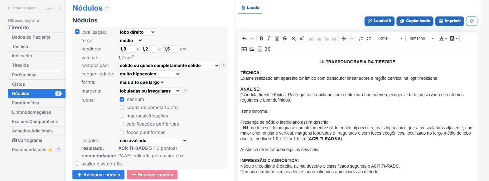
This is exactly where Laudário comes in as a tool supporting the sonologist's routine. The system offers ready-made thyroid ultrasound templates (with and without Doppler) already structured according to the ACR TI-RADS recommendations, organized in scanning order, with fields for describing the parenchyma, characterizing each nodule individually and assessing the lymph node chains.
The big difference is the automatic TI-RADS classification integrated into the report workflow: as the physician checks the composition, echogenicity, shape, margin and echogenic foci of each nodule, the system calculates the total score and assigns the corresponding TR category, automatically suggesting the FNA or follow-up cutoff according to lesion size. This eliminates mental calculation under pressure and drastically reduces the risk of inconsistent classifications between reports. Calculations such as thyroid volume are also done within the typing workflow, with no manual math.
Phrases and conclusions are fully customizable, allowing each professional to adjust the text to their own reporting style without giving up standardization and consistent terminology across exams from the same clinic. The patient history is centralized, which makes comparison with prior reports easier at follow-up, and the final version can be printed with a QR Code for digital validation, adding security and traceability to the document delivered to the patient. The library also covers breast, axilla, abdomen, pelvis, obstetrics and Doppler, all in a single online system.
If you want to reduce rework, standardize TI-RADS stratification and save time in your thyroid routine, Laudário is worth getting to know. The trial is free for 15 days, with no credit card, and the system already comes with the complete library of ultrasound templates ready to use.
The ultrasound reporting platform built by sonologists. We write about reports, classifications and standardization from the real routine of the practice. Who writes and how.
Educational content for healthcare professionals. It does not replace clinical judgment or the guidelines adopted by your service. Editorial policy.