BI-RADS in breast ultrasound: how to describe lesions and standardize the report
How to describe breast lesions on ultrasound using the BI-RADS lexicon (shape, orientation, margin, echo pattern and posterior features) and close the report with a category from 0 to 6 and the corresponding management.
 LaudárioUltrasound reports in practice
This article is part of the guide Standardized classifications in the report: a guide to BI-RADS, TI-RADS and O-RADS.
In breast ultrasound, BI-RADS (Breast Imaging Reporting and Data System), from the American College of Radiology, is the system that standardizes how a finding is described, what risk of malignancy is assigned to it and what management is expected. In practice, describing a lesion with BI-RADS means recording, for each focal finding, the lexicon descriptors (shape, orientation, margin, echo pattern, posterior acoustic features, surrounding tissue and vascularity), its precise location and its dimensions in three planes, and then closing the report with a category from 0 to 6 directly supported by that description.
The final category summarizes management: 0 is incomplete assessment (needs additional evaluation), 1 is a negative exam, 2 are typically benign findings, 3 are probably benign findings (risk of malignancy below 2%, short-interval follow-up, usually 6 months), 4 are suspicious findings with an indication for biopsy (4A low suspicion, 4B intermediate suspicion, 4C moderate suspicion), 5 are findings highly suggestive of malignancy (probability ≥ 95%) and 6 is malignancy already confirmed by biopsy. The most common error is not choosing the wrong category: it is concluding “BI-RADS 3” or “BI-RADS 4A” when the body of the report does not provide the attributes that justify that category. Standardizing the lexicon is what makes the report comparable across exams and across professionals.
Why standardize the breast ultrasound report with BI-RADS
Breast ultrasound is no longer just an adjunct to mammography and has taken a central role in screening routines, especially in women with dense breasts, young women, pregnant women or those being followed for already characterized lesions. With the growing indications (supplemental screening, evaluation of palpable findings, post-biopsy follow-up and correlation with MRI), the way a finding is described and categorized has a direct impact on clinical management, the decision to biopsy and follow-up. And it is precisely in this exam, where variability between professionals is high, that standardization makes a difference.
The BI-RADS system, updated in the 5th edition (with a 2025 revision), was created to solve the biggest problem of breast reports: subjective descriptions, inconsistent conclusions and categories that did not translate between services. Even among experienced professionals, the same nodule may be described as “probably benign solid nodule” by one and “nodule with partially defined contours” by another, creating clinical noise and unnecessary anxiety for the patient. The main problems caused by the lack of standardization are:
- Inconsistent lexicon: terms such as “hypoechoic nodule, regular contours” and “well-defined solid nodular image” describe the same thing, but cause confusion when the exam is compared with a previous one.
- BI-RADS category without criteria: the report concludes “BI-RADS 3” without the description providing the attributes that justify that category.
- Missing or generic management: the category is given, but the report does not guide the expected follow-up (6-month follow-up, biopsy, additional mammography).
- No comparison with prior exams: in a screening and follow-up routine, failing to cite the previous exam means losing the most useful information for the treating physician.
- Diagnostic impression disconnected from the body of the report: the description suggests a suspicious lesion, but the conclusion says BI-RADS 2, or vice versa.
Recommended structure for the breast ultrasound report
A good structure follows the logic of the exam: contextualize the indication, assess the parenchyma bilaterally, describe focal findings with the BI-RADS lexicon, assess the axillary lymph node chains and close with a category correlated with the text.
1. Identification, indication and technique
Include the type of exam (screening, diagnostic, follow-up), age, menstrual cycle phase when relevant, pertinent clinical data (mastalgia, palpable lump, discharge, family history) and any technical limitations (implants, scars, very large breasts). This block gives context to the referring physician and protects the sonologist in technically limited exams.
2. Breast composition and parenchyma
Describe the parenchymal composition (homogeneous fibroglandular, heterogeneous fibroglandular or predominantly fatty) and the presence of diffuse changes, such as ductal ectasia, simple cysts or fibrocystic changes. Commenting on the symmetry between the breasts and the background pattern helps the referring physician interpret the setting in which the focal finding sits.
3. Focal findings with the BI-RADS lexicon
Each lesion should get its own paragraph, with standardized location (breast, quadrant or clock-face position, distance in centimeters from the nipple, depth), dimensions in three planes and the BI-RADS ultrasound lexicon descriptors:
- Shape: oval, round or irregular.
- Orientation: parallel to the skin (more associated with benignity) or not parallel (“taller than wide”, more suspicious).
- Margin: circumscribed or not circumscribed (indistinct, angular, microlobulated or spiculated).
- Echo pattern: anechoic, hyperechoic, complex cystic and solid, hypoechoic, isoechoic or heterogeneous.
- Posterior acoustic features: no posterior features, enhancement, acoustic shadowing or combined pattern.
- Surrounding tissue: architectural distortion, skin thickening, edema, ductal changes.
- Doppler vascularity: absent, internal or peripheral, when applicable.
For microcalcifications seen on ultrasound, describe them and correlate with mammography whenever available, avoiding a conclusion based on the sonographic image alone.
4. Axillary assessment
Even in exams focused on the breasts, assessment of the axillary lymph node chains is a natural part of the study. Describe the presence of lymph nodes, morphology (thin cortex and preserved fatty hilum vs. thickened cortex and loss of the hilum), dimensions of the most representative nodes and Doppler vascularity pattern, when indicated. In oncologic patients or those with suspicious lesions, this block carries even more weight.
5. BI-RADS categorization and suggested management
The conclusion should present, in a hierarchical way, the final BI-RADS category, always aligned with the descriptive body. The current standard recognizes the following categories:
| Category | Meaning | Management |
|---|---|---|
| BI-RADS 0 | Incomplete assessment | Additional evaluation with mammography, MRI or comparison with prior exams |
| BI-RADS 1 | Negative exam, no findings | Routine screening according to age |
| BI-RADS 2 | Typically benign findings (simple cysts, typical intramammary lymph nodes, intact implants) | Routine, no additional follow-up needed |
| BI-RADS 3 | Probably benign findings (risk of malignancy below 2%) | Short-interval ultrasound follow-up, usually 6 months |
| BI-RADS 4 | Suspicious findings. Subdivided into 4A (low suspicion), 4B (intermediate suspicion) and 4C (moderate suspicion) | Biopsy |
| BI-RADS 5 | Findings highly suggestive of malignancy (probability ≥ 95%) | Biopsy and oncologic workup / treatment planning |
| BI-RADS 6 | Malignancy already confirmed by biopsy | Pre- or post-treatment follow-up |
Keeping these criteria always in the same terms, and citing them in the body of the report, is what gives follow-up its consistency. In patients with increased risk factors (family history, known genetic mutations, personal history of a high-risk lesion), it is worth explicitly mentioning these clinical data when provided in the request, because they change the interpretation of the same category.
Common errors in the breast ultrasound report
Even with a defined structure, some recurring errors undermine the quality of breast reports:
- Categorizing without describing the criteria: citing only “BI-RADS 3” or “BI-RADS 4A” in the conclusion when the lesion paragraph does not give the shape, orientation, margin, echo pattern and posterior features that justify the category.
- Mixing a personal lexicon with the BI-RADS lexicon: alternating between “regular contours” and “circumscribed margins” within the same report, or between “well-delimited hypoechoic nodule” and “oval solid nodule parallel to the skin” for the same finding.
- Imprecise location: describing only “upper outer quadrant of the right breast” without clock-face position and distance from the nipple makes correlation with biopsies and future exams difficult.
- Not comparing with prior exams: failing to cite the previous ultrasound when a BI-RADS 3 lesion is under follow-up means losing the most useful information for the treating physician.
- Final category disconnected from the description: stating “BI-RADS 4A” in the impression when the body of the report describes an oval, parallel nodule with circumscribed margins and posterior acoustic enhancement, characteristic of BI-RADS 3, or vice versa.
- Choosing a more conservative category “to be safe”: overestimating a lesion leads to unnecessary biopsies; underestimating delays the diagnosis of a neoplasm. The category must follow the descriptors, not the discomfort.
- Forgetting the axillae: closing the report without citing the assessment of the lymph node chains, especially in patients with a BI-RADS 4 or 5 lesion.
How Laudário helps structure breast reports
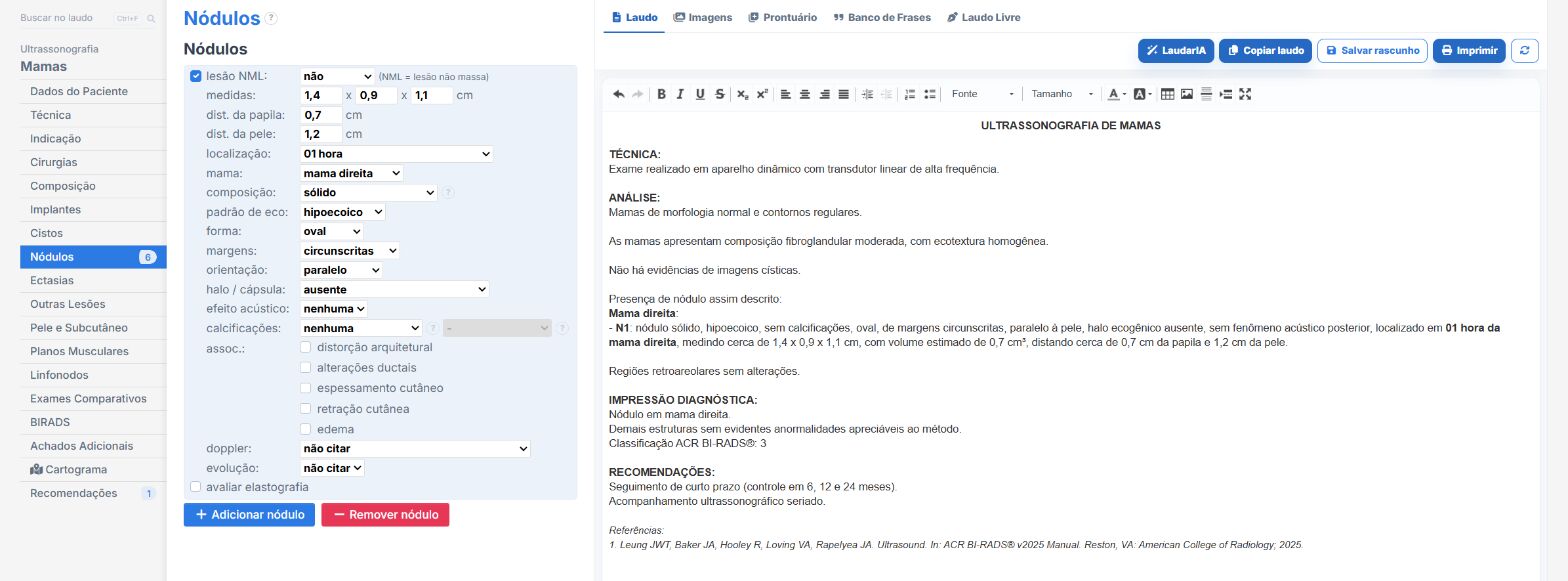
Laudário was designed for professionals who face this routine every day. For breast ultrasound, and for the main protocols of the specialty, the system offers:
- Ready-made, structured templates for breast, breast with Doppler, breast and axilla, and breast and axilla with Doppler, organized in the classic exam sequence to speed up typing.
- Standardized, customizable BI-RADS lexicon to describe shape, orientation, margin, echo pattern, posterior features and surrounding tissue, keeping terminology consistent across reports and professionals in the same clinic.
- Automatic BI-RADS classification (v2025) integrated into the typing workflow on the Premium plan: you select the descriptors and the system applies the corresponding category, reducing classification errors and disconnection between description and impression.
- Calculator and structures for location (quadrant, clock-face position, distance from the nipple), ensuring a precise description that is comparable between exams.
- Complete library with a TI-RADS calculator for the thyroid, obstetric Doppler calculators, fetal percentiles and templates for more than 30 exam types, covering breast, axilla, thyroid, small parts, abdomen, pelvis, obstetrics and Doppler in a single online system.
The result is simple: less time typing, fewer forgotten descriptors and more consistent reports across exams of the same patient, which makes all the difference in a screening and follow-up routine, where semiannual or annual ultrasound follow-up is a central part of management. Instead of starting from a blank page for every patient, you adjust an already tested template, maintain the clinic's professional standard and focus on what really requires your attention: reading the images and making the clinical decision.
If you still assemble your breast reports manually or by copying old texts, try it free for 15 days and see in practice how a well-designed structure, with BI-RADS classification built into the workflow, saves time on every exam.
The ultrasound reporting platform built by sonologists. We write about reports, classifications and standardization from the real routine of the practice. Who writes and how.
Educational content for healthcare professionals. It does not replace clinical judgment or the guidelines adopted by your service. Editorial policy.