Axillae and lymph nodes: how to structure the axillary staging report
Size is not a criterion: metastasis starts in the cortex. See the 3 mm that decide the study, why the hilum is the strongest axis, the Berg levels, and why an axilla that looks normal on ultrasound does not replace sentinel node biopsy.
 LaudárioUltrasound reports in practice
This article is part of the guide Standardized classifications in the report: a guide to BI-RADS, TI-RADS and O-RADS.
In breast cancer, axillary status is the single most important prognostic factor, and ultrasound is the only routine method able to look at it before surgery, with enough resolution to measure millimeters of cortex and even to biopsy the suspicious node in the same session. Few studies combine this much access and consequence.
Even so, the axilla usually appears in the breast report as a footer sentence: "axillary lymph nodes of usual appearance". No measurement, no level, no description of the cortex. This text treats the axilla as the study it is: the morphologic criteria that separate reactive from suspicious, the number that decides everything, the anatomic levels and what the surgeon does with this information.
The normal lymph node and why it is easy to recognize
The normal lymph node has a characteristic and reassuring sonographic anatomy:
- Reniform shape (oval, flattened): long axis clearly greater than the short axis.
- Thin, homogeneous cortex, hypoechoic, surrounding…
- …the central fatty hilum, hyperechoic, continuous with the adjacent tissue.
- Hilar vascularity on Doppler: the vessels enter through the hilum and branch from it, centrifugally.
A detail that prevents many unnecessary reports: size is not a criterion. A 3 cm lymph node with a thin cortex, preserved hilum and hilar vascularity is normal, just large. And an 8 mm lymph node with a 4 mm cortex and an effaced hilum is suspicious. Metastatic disease starts in the cortex, in the subcapsular sinus, and that is where it must be sought. Reporting "lymphadenopathy" by size, without describing the cortex, is measuring the wrong thing.
The 3 mm: the most important number in the study
Cortical thickness is the single parameter with the best performance for suspicion, and the cutoffs used in practice are:
- Up to 3 mm: within expected limits.
- Above 3 mm: atypia; deserves detailed description and, depending on the context, biopsy.
- 4 mm or more: high suspicion.
Two technical points on how to measure, because they change the number:
- Measure perpendicular to the hilum, at the point of greatest thickness of the cortex: not the average, not any point. Early metastatic thickening is focal and eccentric; measuring at a spared point gives a false negative.
- Measure without compression. The transducer flattens the cortex easily.
And there is a morphologic distinction worth more than the millimeter: diffuse, homogeneous thickening suggests reaction; focal, eccentric or lobulated thickening suggests metastasis. A uniform 3.5 mm cortex in a patient with dermatitis of the arm is something different from a 3.5 mm cortex bulging in a single quadrant, in a patient with an ipsilateral BI-RADS 5 nodule.
Reactive or suspicious: the three axes
The reading is organized along three axes, and the strongest of them is the hilum.
1. Cortex
- Reactive: diffuse and homogeneous, concentric thickening.
- Suspicious: focal, eccentric or lobulated thickening; heterogeneous cortex.
2. Hilum: the decisive axis
- Reactive: preserved hilum, even with a thick cortex. The inflamed node swells, but keeps its architecture.
- Suspicious: hilum displaced to the periphery (the metastasis grows eccentrically and pushes) and, more serious, effaced hilum, replaced by tumor.
If there is one finding to take from this text, it is this: the reactive node preserves the hilum; metastasis destroys it. A thick cortex with an intact hilum is almost always reaction. An effaced hilum is suspicious even with a modest cortex.
3. Vascularity on Doppler
- Reactive: hilar, symmetric flow, branching from the center outward: even when increased, the pattern is maintained.
- Suspicious: subcapsular and peripheral flow (the metastasis brings its own neovascularization through the capsule), disorganized pattern, or absence of hilar flow.
The vascular pattern is what resolves the cases in which the morphology is ambiguous: peripheral flow in a node of nearly normal morphology is an early and underappreciated sign.
The remaining findings
- Rounding: long axis/short axis ratio below 2. The node that loses its reniform shape and becomes spherical.
- Intranodal microcalcifications: rare, but highly specific; associated with ductal carcinoma with a calcified component.
- Intranodal cystic areas: necrosis. A high-suspicion finding.
- Loss of margins / extracapsular extension: advanced disease, with direct surgical implications.
The Berg levels: where the node is matters
The axilla is not a single compartment, and a report that does not state the level delivers half the information. The anatomic reference is the pectoralis minor muscle:
- Level I: lateral to the border of the pectoralis minor. It is the level of primary drainage of the breast and where the sentinel node lives in the overwhelming majority of cases. Most metastases start here.
- Level II: posterior to the pectoralis minor, including the Rotter (interpectoral) nodes.
- Level III: medial to the border of the pectoralis minor, infraclavicular. Involvement here already signals more advanced disease.
Beyond the axilla proper, two chains need to be scanned and are almost always forgotten:
- Internal mammary chain: in the parasternal intercostal spaces. Alternative drainage, particularly relevant in medial and central tumors. Involvement changes the staging and the radiotherapy field.
- Supraclavicular chain: involvement here is advanced regional disease.
A single suspicious node at level I is one clinical story; the same node with a second at level III is a completely different one. The level is staging information, and it is free: just place the transducer on the pectoralis minor and orient yourself by it.
What the surgeon does with this report
Understanding the downstream decision explains why the details matter.
The standard management in the clinically negative axilla is sentinel lymph node biopsy: the first draining node is removed and, if it is negative, the axilla is spared, avoiding axillary dissection and the lymphedema that accompanies it. It is one of the great achievements of breast surgery in recent decades.
Ultrasound comes in before that, and the objective is precise: to identify the patient whose axilla is already positive, to take her straight to biopsy. A suspicious node with a positive FNA or core biopsy confirms axillary disease without diagnostic surgery and repositions all of the planning, including the discussion of neoadjuvant chemotherapy.
Hence the editorial consequence: a report that describes a suspicious node should suggest biopsy. Describing "atypical axillary lymphadenopathy" and stopping there leaves the finding without an outcome. And when the biopsy is positive and the patient goes to neoadjuvant therapy, node clip marking comes in, which is also an ultrasound-guided procedure.
An honest note on calibration: axillary ultrasound has high specificity and limited sensitivity. It does not detect micrometastasis, and an axilla that is normal on ultrasound does not replace sentinel node biopsy. The study serves to find who is already positive, not to clear who looks negative. A report that suggests otherwise induces error.
BI-RADS and the lymph node
The relationship is simpler than one might imagine, and binary in practice: an atypical lymph node is BI-RADS 4; typical or reactive, BI-RADS 2. There is no 3, there is no 4A/4B/4C subdivision for lymph nodes. The logic is the same as in the rest of the system: the category points to management, and for an atypical node the management is tissue.
If you want the reasoning of the complete system, it is in BI-RADS in breast ultrasound.
Accessory breast: the finding that frightens for no reason
Ectopic breast tissue in the axilla is common, congenital and benign, a remnant of the embryonic milk line. On ultrasound, it is tissue with echotexture identical to the breast parenchyma, without a capsule, without a hilum, located in the axillary subcutaneous tissue.
It matters for two opposite reasons. First, it is mistaken for lymphadenopathy and for lipoma, generating unnecessary workup; the glandular echotexture resolves it. Second, and more relevant: accessory breast is breast tissue, and breast tissue gets sick. It can become engorged in pregnancy and lactation (the classic cause of the "axillary lump that appeared postpartum"), it can have a fibroadenoma and it can have carcinoma. Describing it as accessory breast is also saying that it deserves to be examined with the same lexicon as the breast.
Pitfalls of the axillary report
- Using size as the criterion: the disease starts in the cortex; a large node with a preserved hilum is normal.
- Not measuring cortical thickness: it is the number that decides.
- Measuring the cortex at a spared point: early thickening is focal; measure at the thickest point.
- Compressing with the transducer: it flattens the cortex and underestimates.
- Ignoring the hilum: it is the strongest axis; reactive preserves, metastasis effaces.
- Not describing the vascular pattern: peripheral flow is an early sign.
- Not stating the level: I, II and III have distinct prognostic meanings.
- Forgetting the internal mammary and supraclavicular chains: they change the staging.
- Describing a suspicious node without suggesting biopsy: it leaves the finding without an outcome.
- Suggesting that a normal axilla replaces the sentinel node: the sensitivity does not allow it.
- Confusing accessory breast with lymphadenopathy, and vice versa.
How Laudário helps structure the axillary report
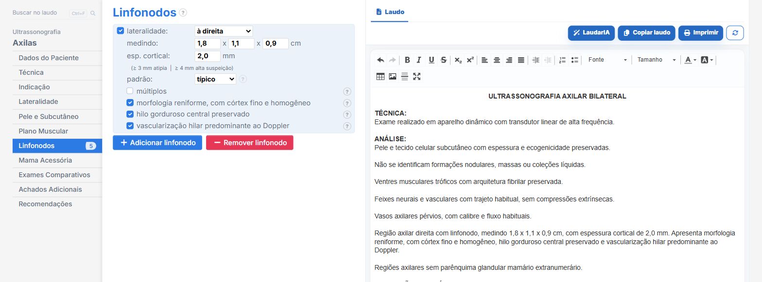
Laudário has a dedicated Axillae module, in addition to Breasts and Axillae (with and without Doppler) and General Lymph Node for the other territories. In practice:
- A cortical thickness field with the inline reference (above 3 mm atypia; 4 mm or more high suspicion) and automatic reclassification to the atypical pattern when the entered value crosses the threshold, so the number does not go unnoticed in the rush.
- The three patterns (typical, reactive, atypical) with their own subitems already written, and the twelve descriptors of atypia: rounding with the L/S ratio, focal/diffuse/lobulated thickening, displaced or effaced hilum, absence of hilar flow, subcapsular flow, disorganized pattern, necrosis, microcalcifications.
- In the Breast report, chains by level (I, II and III), internal mammary and supraclavicular as their own locations, with the intramammary node positioned by clock face.
- Lymph node BI-RADS derived automatically from the pattern: atypical 4, typical or reactive 2.
- Auto-suggested biopsy recommendation whenever there is an atypical node, so the finding never goes without an outcome.
- Accessory breast as its own field, with laterality and a ready-made phrase.
- Modules for guided FNA and core biopsy, and for breast nodule clip placement, for when the report becomes a procedure.
- Support from LaudarIA for multichain cases and post-neoadjuvant follow-up.
If the axilla is still a footer sentence in your breast reports, it is worth trying: 15-day free trial, no credit card, with all breast modules unlocked.
The ultrasound reporting platform built by sonologists. We write about reports, classifications and standardization from the real routine of the practice. Who writes and how.
Educational content for healthcare professionals. It does not replace clinical judgment or the guidelines adopted by your service. Editorial policy.