Ultrasound-guided breast core biopsy and clip placement: what to record in the report
Indications for ultrasound-guided percutaneous core needle biopsy, when to place the metallic clip, how to document the needle in the lesion, the number of cores, radiologic-pathologic correlation, and what the procedure report needs to contain.
 LaudárioUltrasound reports in practice
This article is part of the guide Standardized classifications in the report: a guide to BI-RADS, TI-RADS and O-RADS.
Ultrasound-guided percutaneous core needle biopsy (core biopsy) is the method of choice for investigating suspicious breast nodules. The procedure report is a clinical document and also a quality record: it must allow another physician to know exactly which lesion was biopsied, how, and whether the pathology result makes sense with the imaging.
Indications
- BI-RADS 4 and 5 lesions visible on ultrasound.
- Some BI-RADS 3 lesions, when there is a clinical indication (patient preference, inability to follow up, pregnancy or surgery planning).
- Suspicious axillary lymph nodes, for staging.
Technique and documentation
- Consent, asepsis, and local anesthesia.
- Large-gauge needle (generally 14G), with a path as parallel to the chest wall as possible.
- Images documenting the needle before and after firing, crossing the lesion, preferably in two planes.
- Enough cores for diagnosis, generally three to five, from different regions of the lesion.
- Assessment of the cores (whether they sink in saline, whether they are intact) as an indicator of an adequate sample.
When to place the clip
The metallic clip marks the site of the lesion so it can be found later. It is indicated when:
- The lesion is small and may no longer be visible after the biopsy.
- The patient is going to receive neoadjuvant chemotherapy, which may make the lesion disappear.
- There are several lesions and it is necessary to identify which one was biopsied.
After placement, the clip position is checked on ultrasound and, generally, on a mammogram, because the clip can migrate. The distance between the clip and the lesion should be recorded.
Radiologic-pathologic correlation
The pathology result must be concordant with the imaging. A BI-RADS 5 nodule with a benign result is discordant and requires repeat biopsy or excision. This step is the responsibility of whoever performed the biopsy and should appear in the follow-up.
What the procedure report should include
- Indication and BI-RADS category of the lesion.
- Location of the lesion (side, quadrant or clock-face position, distance from the nipple) and measurements.
- Consent, asepsis, and anesthesia.
- Needle gauge, number of cores, and access route.
- Confirmation of the needle in the lesion.
- Clip: whether it was placed, type, and final position.
- Complications (hematoma, bleeding, vasovagal reaction) and instructions.
- Recommendation for correlation with the pathology result.
Common errors
- Not recording the clock-face position and the distance from the nipple.
- Missing the image of the needle crossing the lesion.
- Not stating the number of cores.
- Placing a clip without checking and recording the final position.
- Accepting a discordant benign result without recommending a new approach.
See also our text on BI-RADS in breast ultrasound.
How Laudário helps
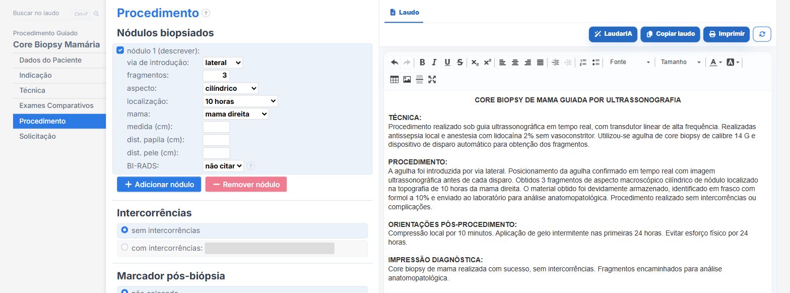
In the core biopsy and breast nodule clip placement templates of Laudário:
- Target lesion with location, measurements, and BI-RADS category.
- Needle, number of cores, clip, and complications in their own fields.
- Instructions and recommendations in their own tab.
Try it for 15 days free, no credit card required.
The ultrasound reporting platform built by sonologists. We write about reports, classifications and standardization from the real routine of the practice. Who writes and how.
Educational content for healthcare professionals. It does not replace clinical judgment or the guidelines adopted by your service. Editorial policy.