Nodule elastography: breast, thyroid and prostate, from the Itoh score to shear wave
Elastography is not one exam: it is two techniques with different scales and units. See the Itoh score, the strain ratio, why kPa and m/s cannot be converted by simple proportion, and how stiffness moves a BI-RADS 3 to 4 without ever touching a 5.
 LaudárioUltrasound reports in practice
This article is part of the guide Standardized classifications in the report: a guide to BI-RADS, TI-RADS and O-RADS.
Elastography gave ultrasound a piece of information that the physician has always had and the machine never saw: stiffness. Palpating a nodule and feeling it stony hard is a centuries-old clinical datum; elastography turns that datum into an image and, in some modes, into a number. The promise is straightforward: a malignant lesion tends to be stiffer than the surrounding tissue.
The promise is real, but it comes with fine print that decides whether the exam helps or hinders. Elastography is not one exam: it is two different physical techniques, with different scales, different cutoffs by organ and units that cannot be converted by simple proportion. And above all, it is a complementary datum: it adjusts a BI-RADS or TI-RADS category at the margin, it does not replace it. This text walks through all of that.
Two techniques, two types of response
The fundamental distinction, and the source of most of the confusion:
| Strain (compression) | Shear wave | |
|---|---|---|
| What it measures | Relative deformation | Wave velocity, giving absolute stiffness |
| Stimulus | Transducer compression (or the body's own pulsation) | Acoustic pulse from the machine (ARFI) |
| Response | Qualitative (color map) + semiquantitative (ratio) | Quantitative (m/s or kPa) |
| Operator dependent | Yes, very | Little |
| Needs a reference | Yes: adjacent normal tissue | No |
The practical consequence is large. Strain says that this lesion is stiffer than that tissue next to it: it is comparative, and so without adjacent normal tissue it simply does not work. Shear wave gives a number that exists on its own and is comparable between exams and, with caveats, between machines.
Itoh score: the qualitative scale (1 to 5)
The Tsukuba scale, or Itoh score, reads the strain color map and classifies the deformation pattern. It runs from 1 to 5, from elastic to stony hard:
- 1: homogeneous deformation throughout the lesion. Predominantly elastic pattern.
- 2: deformation in most of the lesion, with small focal areas of greater stiffness.
- 3: deformation restricted to the periphery, with stiffness of the central portion. Heterogeneous pattern.
- 4: no deformation across the entire extent of the lesion. Global stiffness.
- 5: no deformation in the lesion and in the adjacent tissue. Marked stiffness.
The practical reading: 1 and 2 point to benignity; 4 and 5 point to suspicion; 3 is genuinely indeterminate, and this is where maturity shows. A score of 3 should not be pushed to one side for convenience. A report that states "indeterminate elastographic behavior" is more useful than one that pretends to decide.
Note the jump from 4 to 5: what changes is not the lesion, it is the surroundings. A score of 5 means that the stiffness extends beyond the limits of the lesion, the elastographic correlate of desmoplastic reaction, and therefore the most concerning.
Strain ratio: the number of strain
The strain ratio compares the deformation of the lesion with that of an area of normal tissue at the same depth. It is semiquantitative: a number, but dependent on the reference chosen.
Cutoffs vary by organ and by study; in practice, values around 3.0 for thyroid and 3.5 for breast are among the most used. Above the cutoff, suspicion.
Three situations make the strain ratio not calculable, and the report should say so instead of inventing a value:
- Absence of adjacent normal parenchyma: without a reference, there is no ratio.
- Multiple nodules occupying the field, with no normal tissue available.
- Bulky lesion, which leaves no comparable area in the same plane.
This is a structural limitation of the method, not an operator failure, and it is exactly why shear wave exists.
kPa and m/s: the conversion that is not a simple proportion
This is the most important technical point of the text, and the one most often gotten wrong.
Shear wave measures the velocity of the shear wave (c, in m/s). The machine converts this into elastic modulus (E, in kPa) through a relationship that is quadratic, not linear:
E ≈ 3ρc²
Since the density of soft tissue is close to 1 g/cm³, this reduces in practice to:
E (kPa) ≈ 3 × c² and c (m/s) ≈ √(E / 3)
A few pairs, to calibrate intuition:
| Velocity (m/s) | 2.0 | 2.5 | 3.0 | 4.0 | 4.7 | 5.2 |
|---|---|---|---|---|---|---|
| Stiffness (kPa) | 12 | 19 | 27 | 48 | 66 | 81 |
Note what the curve does: doubling the velocity quadruples the stiffness. This is why a cutoff validated in m/s cannot be carried over to kPa "proportionally", and vice versa. A cutoff of 80 kPa corresponds to about 5.2 m/s, not 2.9. A cutoff of 2.5 m/s corresponds to about 19 kPa, not 65.
The practical rule that avoids the error: use the cutoff in the unit in which it was validated, and set the machine to that unit. If your practice works in kPa, adopt cutoffs published in kPa. Converting by hand, in the middle of the exam, is how the method fails silently: the number comes out plausible and is wrong.
And an honest caveat: cutoffs vary by manufacturer, by organ and by study. There is no universal number. It is the same problem that liver elastography faces, and the solution is the same: know the cutoffs of your equipment and keep them stable throughout follow-up.
How the parameters talk to each other: the voting system
A robust way to organize the conclusion is to treat each parameter as a vote, instead of forcing a single score:
- Itoh ≤ 2 votes benign; ≥ 4 votes suspicious; 3 does not vote.
- Strain ratio below the cutoff votes benign; above it, suspicious.
- Shear wave below the threshold votes benign; above it, suspicious.
If all vote the same way, the conclusion is straightforward. If they vote on opposite sides, the result has a name of its own: discordant elastographic parameters. Reporting the discordance as a category is more honest, and more useful, than arbitrarily choosing a winner. A lesion with Itoh 4 and a strain ratio of 1.8 is a lesion on which elastography has not made up its mind, and the clinician needs to know that.
Integration with BI-RADS and TI-RADS: conservative and asymmetric
This is the question everyone asks: does elastography change the category? The answer is yes, but little, and deliberately conservatively:
- Stiffness raises 3 → 4. A nodule morphologically consistent with BI-RADS 3 that proves stiff goes up to 4 and now deserves biopsy.
- Low stiffness lowers 4 → 3. A nodule only mildly suspicious on morphology that is frankly soft may go back to 3 and follow-up.
- Never touches 5. A lesion with classic morphology for malignancy is not rescued by soft elastography. And, in practice, this extends to 4B and 4C: the higher the morphological category, the less elastography should move it.
The asymmetry is intentional and reflects the cost of errors: raising a category costs a biopsy; lowering a category may cost a diagnosis. That is why downgrading is the most dangerous move, and it should be reserved for lesions whose morphological suspicion was already marginal.
The corollary is what matters most: elastography does not replace the morphological lexicon. Shape, orientation, margin, echo pattern and associated findings continue to decide. If you do not yet use these systems in a structured way, start with them: the guide is at Standardized classifications in the report: BI-RADS, TI-RADS and O-RADS, and the details are in BI-RADS in breast ultrasound and Thyroid ultrasound and TI-RADS.
Each organ has its own yardstick
Breast
This is where the evidence is most robust and the gain clearest: elastography improves specificity, reducing biopsies of benign BI-RADS 3 and 4A lesions. The most defensible context of use is exactly this: complementing a BI-RADS 3 or 4A lesion. Beware of fibroadenoma, which can be stiff, and of mucinous carcinoma, which can be soft: these are the classic exceptions that keep elastography from deciding alone.
Thyroid
Real gain in complementing the indeterminate nodule and in screening within multinodular goiter: which nodule to puncture when there are eight? One specific pitfall: coarse calcification and intense fibrous component stiffen without malignancy, and predominantly cystic nodules have nothing to deform. Elastography of a cystic nodule means nothing.
Prostate
Transrectal, in the setting of elevated PSA, it helps direct the biopsy to the stiffest focus of the peripheral zone. It is an adjunct to biopsy, not a substitute, and here there is no consolidated shear wave threshold as there is in the breast.
Soft tissues
The least standardized territory: there are no established cutoffs, and elastography enters as additional characterization of an indeterminate lesion, without decision-making power of its own.
The limitations that must be in the report
Elastography has failure modes that the color map does not reveal. When present, they belong in the document:
- Depth: deep lesions receive less energy and the measurement degrades.
- Motion artifact: breathing, pulsation, unsteady hand.
- Calcifications and intense fibrosis: they stiffen without malignancy.
- Cystic component: fluid does not deform like a solid; on strain it generates the classic three-layer artifact, which is noise, not stiffness.
- Bulky lesion: no reference for the ratio.
- Excessive compression on strain: it artificially stiffens everything. The technique calls for minimal pressure.
- Inadequate elastographic window: without correlation between the B-mode plane and the elastographic map plane, the measurement is not of the lesion.
Pitfalls of the nodule elastography report
- Converting kPa to m/s by simple proportion: the relationship is quadratic; the number comes out plausible and wrong.
- Using one manufacturer's cutoff on another's value.
- Forcing Itoh 3 to one side: indeterminate is a legitimate answer.
- Performing elastography of a cystic nodule: there is nothing to deform.
- Downgrading a BI-RADS 5 because of softness: morphology rules.
- Compressing too much on strain: it stiffens everything and produces false positives.
- Calculating strain ratio without adjacent normal parenchyma: report "not calculable" and say why.
- Reporting stiffness without the technique used: strain and shear wave are not the same datum.
- Omitting the technical limitation: depth, calcification and motion change the value.
- Treating elastography as a verdict: it adjusts the category at the margin, it does not replace it.
How Laudário helps
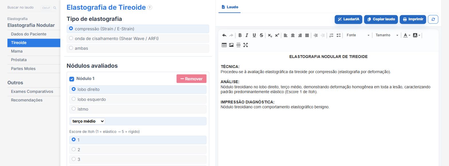
Laudário has a Nodule Elastography module with tabs by organ (thyroid, breast, prostate (transrectal) and soft tissues), each with its own yardstick. In practice:
- Choice of technique (compression, shear wave or both) that reconfigures the interface: only the fields that technique produces appear.
- Itoh score from 1 to 5 with the semiology written out for each level: the report describes the deformation pattern, not just the number.
- Organ-specific cutoffs for strain ratio and shear wave, displayed next to the field, with a selectable unit (kPa or m/s).
- Voting system among Itoh, strain ratio and shear wave, with "discordant" and "indeterminate" as legitimate conclusions instead of forced decisions.
- Strain ratio "not calculable" as its own field, with the three reasons, written into the report.
- Integration with the Breast report: stiffness raises the nodule from category 3 to 4 and softness lowers it from 4 to 3, without ever touching category 5.
- Technical limitations in italics in the report: window, motion, depth, calcification, dimensions.
- Support from LaudarIA for cases in which the parameters contradict each other.
If you already have elastography on your machine and have not yet incorporated it into the report, it is worth trying: 15-day free trial, no credit card required, with all modules unlocked.
The ultrasound reporting platform built by sonologists. We write about reports, classifications and standardization from the real routine of the practice. Who writes and how.
Educational content for healthcare professionals. It does not replace clinical judgment or the guidelines adopted by your service. Editorial policy.