Deep endometriosis mapping: how to structure the report by compartment according to the IDEA consensus
The IDEA consensus organizes endometriosis mapping into four steps and anatomical compartments. See how to report nodules, the sliding sign, the distance from the anal verge and the infiltrated layer, and why preparation defines what the exam can conclude.
 LaudárioUltrasound reports in practice
This article is part of the guide Gynecologic ultrasound: the complete guide, from uterus to ovaries.
Transvaginal ultrasound is no longer an exam that “does not see endometriosis” and has become the first-line method for mapping deep disease, but this is only true when the exam is performed with preparation, with dynamic maneuvers and with a report organized by anatomical compartments. The IDEA consensus (International Deep Endometriosis Analysis group, Guerriero et al., 2016) established the common language of this mapping: four systematic steps that range from the basic assessment of the uterus and adnexa to the targeted search for infiltrative nodules in the anterior and posterior compartments, passing through the soft markers and the sliding sign.
The practical difference is large. A report that concludes “uterus and adnexa unremarkable” in a patient with disabling dysmenorrhea and deep dyspareunia has not merely missed the diagnosis: it has delayed, on average, years of investigation. A report that describes a 1.8 cm retrocervical nodule infiltrating the muscularis propria of the rectum 8 cm from the anal verge, with an obliterated pouch of Douglas and a negative sliding sign, hands the surgeon practically the operative plan. This text walks through the complete structure of that report, compartment by compartment.
Why the compartment-based report changed the diagnosis of endometriosis
Deep infiltrating endometriosis (DIE) is defined by the infiltration of ectopic endometrial tissue more than 5 mm beneath the peritoneal surface. It is a disease of topography: it does not appear diffusely and randomly, but at predictable sites, following the path of retrograde menstrual flow and the areas of lower peritoneal mobility. Precisely because it is predictable, it is traceable, as long as the examiner searches each site in a targeted way.
Before IDEA, each service had its own vocabulary. “Nodule in the cul-de-sac”, “retrocervical thickening”, “posterior adhesion” and “septal lesion” could describe the same thing or completely different things, and the surgeon could not tell which. With the compartment-based taxonomy, the report now always answers the same questions, in the same order: is the anterior compartment involved? And the central? And the posterior? And the lateral? Is the cul-de-sac obliterated? Does the disease reach the bowel, and if so, at what height and in which layer?
This change has a direct consequence in the operating room. Involvement of the posterior compartment with infiltration of the rectal muscularis propria defines the need for a colorectal surgeon on the team. Ureteral involvement defines the need for a urologist and, possibly, prior stenting. A report that does not separate these compartments forces the team to find this out intraoperatively, the worst possible moment.
The four steps of the IDEA consensus
The consensus proposes an exam sequence, and that same sequence should organize the report:
- Step 1: basic assessment of the uterus and ovaries, including the sonographic signs of adenomyosis and the search for endometriomas.
- Step 2: soft markers: site-specific tenderness with the transducer and ovarian mobility.
- Step 3: status of the pouch of Douglas, assessed in real time by the sliding sign.
- Step 4: search for deep endometriosis nodules in the anterior and posterior compartments.
Note what this order reveals: two of the four steps are dynamic. Deep endometriosis is, to a large extent, a diagnosis made on a moving exam, not on a frozen image. A report built only from static images misses half of the consensus.
Step 1: uterus, ovaries and the distinction between endometrioma and adenomyosis
The assessment starts with the conventional, but with two specific points of attention.
The endometrioma has an established description and should be reported with it, not as a “complex cyst”: an ovarian cystic image, typically unilocular, with smooth walls, with fine homogeneous low-level echoes in suspension (the classic ground-glass appearance), without a solid component or papillary projection and without internal vascularity on Doppler. Described this way, it already comes out classified as O-RADS 2 when it measures up to 10 cm. It is worth recording dimensions and volume, because follow-up and the surgical decision depend on them, and because the endometrioma is, on its own, a marker of increased probability of deep disease at other sites. Finding an endometrioma is an invitation to look for more, not an endpoint.
Adenomyosis, on the other hand, needs to appear on a separate line of the conclusion. It is a distinct entity: myometrial involvement, not deep endometriosis. Mixing the two in the same sentence of the diagnostic impression confuses the clinician about what exactly was found. The report can, and should, describe both when they coexist, but as separate findings.
One step 1 sign deserves its own emphasis: kissing ovaries, ovaries adhered to each other in the midline, behind the uterus. It is not an anatomical curiosity. It is a marker of extensive disease, strongly associated with bowel involvement and obliteration of the cul-de-sac, and it should be explicitly cited in the conclusion whenever present.
Step 2: the soft markers that only the dynamic exam reveals
Soft markers are findings that do not appear on any printed image, which is why so many reports omit them. There are two:
- Site-specific tenderness: compression with the transducer reproduces the patient's pain at a specific site. It is worth recording which sites were tested and which were positive: uterine mobilization, right ovary, left ovary, right uterosacral ligament, left uterosacral ligament, rectovaginal septum. It is a pain map that speaks directly to the clinical complaint.
- Ovarian mobility: is the ovary mobile, does it have restricted mobility, or is it fixed? And if fixed, fixed to what: the uterus, the lateral pelvic wall, the sigmoid or the ovarian fossa, close to the ureteral course? An ovary fixed to the ovarian fossa immediately raises the question about the ipsilateral ureter.
These markers matter because adhesive endometriosis can exist without a measurable nodule. A patient with pelvic pain, fixed ovaries and a partially obliterated cul-de-sac has disease, even if no nodular lesion is measured. A report that only describes what can be measured does not describe this patient.
Step 3: the sliding sign and the status of the pouch of Douglas
This is the most specific step of the consensus and the most underused in practice. The sliding sign is a real-time assessment: with the transvaginal transducer and the free hand on the abdominal wall, gentle pressure is applied and one observes whether the peritoneal surfaces slide freely over one another.
There are two distinct planes, and the report should state which one was assessed:
- Retrocervical sliding: does the rectum slide over the posterior face of the cervix and lower third of the uterus?
- Uterine fundus sliding: does the sigmoid slide over the posterior face of the uterine fundus?
The reading is straightforward and defines the conclusion about the pouch of Douglas:
- Positive sliding in both planes: cul-de-sac free, not obliterated.
- Negative sliding in one plane (unilateral): cul-de-sac partially obliterated.
- Negative sliding in both planes (bilateral): cul-de-sac completely obliterated.
A well-constructed report describes this in clinical prose, not jargon: “Dynamic assessment demonstrating a bilateral negative sliding sign, characterizing complete obliteration of the posterior cul-de-sac”. It is also worth recording the level of the obliteration (retrocervical, mid-uterine or fundal), because this anticipates the extent of the surgical dissection.
The same reasoning applies to the anterior compartment, with uterovesical mobility: does the posterior wall of the bladder slide over the anterior wall of the uterus and the cervix? Here there is an important interpretive trap that the report should name: vesicouterine fixation is also caused by prior cesarean scars. Describing “obliterated vesicouterine space” without qualifying whether the probable cause is endometriosis or postcesarean adhesion gives the clinician ambiguous information in a population in which cesarean delivery is extremely common.
Regarding fluid in the cul-de-sac, a note on precision: describing the amount is not enough. The appearance is what informs: anechoic is physiologic; ground-glass suggests blood or endometriosis; with septations or internal adhesions it points to a chronic inflammatory process.
Step 4: the nodules, compartment by compartment
Anterior compartment
It encompasses the bladder, urethra, distal ureters, vesicouterine space and round ligaments.
The bladder requires adequate filling, and the report should state the filling status, because an empty bladder simply does not allow the compartment to be assessed. The most frequent location of the lesion is the posterior wall and the vesicouterine recess, and the data that changes management is the depth of invasion: serosa/subserosa, muscularis (detrusor) without mucosal involvement, or mucosa. A lesion that reaches the detrusor implies partial cystectomy; a lesion that reaches the mucosa changes the approach and often requires prior cystoscopy. Describing only a “1.5 cm bladder nodule” omits exactly the operative data.
The distal ureter is the finding that most silently compromises the patient, because ureteral obstruction can progress to loss of renal function with no symptoms at all. Two pieces of data are mandatory when there is a lesion:
- Nature: extrinsic (external compression or retraction by an adjacent lesion) or intrinsic (infiltration of the ureteral wall itself). The distinction defines whether ureterolysis is enough or segmental resection with reimplantation will be needed.
- Repercussion: presence of stenosis, upstream dilation and visualization of peristalsis. And, in the report, the distance from the ureteral ostium, which guides the surgical approach.
The round ligaments complete the compartment and are often forgotten, especially in the inguinal segment: a patient with a painful nodule in the inguinal region that worsens during the menstrual period may have round ligament endometriosis, and the report rarely considers this.
Posterior compartment
This is the most frequently involved compartment and the most demanding to describe. It involves the uterosacral ligaments, the torus uterinus, the rectovaginal septum, the posterior vaginal wall, the retrouterine recess and the rectosigmoid.
The uterosacral ligaments (USL) should be assessed by segment (insertion at the torus, proximal segment, mid to distal segment), because distal extension brings the lesion closer to the inferior hypogastric plexus and increases the risk of postoperative urinary dysfunction. The typical morphology is hypoechoic plaque-like thickening or a solid hypoechoic nodule, with characteristically irregular margins.
The torus uterinus, the transverse peritoneal band that joins the two USL in the retrocervical region, is the central site. When normal, the retrocervical peritoneal line is intact. When involved, the report should record not only the measurements but also the extent: does it involve the posterior vaginal fornix? Does it extend laterally to the USL? Is it adhered to the anterior wall of the rectum? Since the USL and torus are the same anatomical region, when both are normal it makes sense to describe them in a single sentence, without artificially fragmenting the text.
The rectovaginal septum and the posterior vaginal wall require the distance from the anal verge in centimeters, which is the data the colorectal surgeon uses to decide the technique. And there is a finding with its own name worth knowing: the diabolo nodule (or hourglass nodule), in which the lesion has a component in the posterior vaginal fornix in direct continuity with infiltration of the anterior rectal wall, constricted in the middle. This is the configuration that defines the need for combined resection.
Lateral compartment
Less cited in the original consensus papers, but clinically decisive. It involves the parametria, the parametrial segment of the ureter and the uterine arteries. Parametrial involvement is what brings the disease close to the hypogastric nerve and the sacral plexus, and is the reason why some patients develop bladder dysfunction and neuropathic pain after extensive surgery. When a spiculated nodule is identified in the parametrium, describing whether it lies along the course of these nerves is information the surgeon has no other way of obtaining before surgery.
The bowel compartment: height and layer are everything
Bowel infiltration is the finding that most changes the scale of the surgery, and the report needs to answer three questions with numbers.
Where? Anatomical segmentation is done by distance from the anal margin, and it is worth reporting both the name of the segment and the measured distance:
- Low rectum: 0 to 5 cm from the anal verge
- Mid rectum: more than 5 to 10 cm
- High rectum: more than 10 to 15 cm
- Rectosigmoid junction: more than 15 to 20 cm
- Sigmoid: above 20 cm
Height matters because lesions in the low rectum carry a significantly higher risk of fistula and of needing a protective stoma. A report that says “rectal lesion” without the distance leaves that conversation for later.
How deep? Depth follows the layers of the wall: serosa/peritoneal, muscularis propria, submucosa, mucosa. Infiltration of the muscularis propria is the watershed: below it, the lesion can be treated by shaving; from that point on, discoid resection and segmental resection come into discussion.
How much? Percentage of the circumference involved and degree of luminal stenosis. These two numbers speak to each other: involvement above 50% of the circumference or narrowing of 75% or more are situations that characterize significant luminal stenosis and that tend to tip the decision toward segmental resection. When the lesion is extensive and the patient has cyclic obstructive symptoms, these percentages are what support the indication.
The classic sonographic signs of bowel involvement
Bowel endometriosis has its own iconography, with names that describe exactly what is seen. Using them in the report communicates much more than “wall thickening”:
- Comet sign: infiltrative hypoechoic thickening with progressive narrowing, tapering toward the ends like the tail of a comet.
- Indian headdress: nodular hypoechoic thickening with prominent spicules projecting toward the lumen.
- Mushroom cap: nodule with wall retraction, in which the base infiltrates the muscularis and the top widens, overestimating the real thickness.
- Spiculated plaque: plaque-like thickening with a spiculated contour, typical of the rectosigmoid junction.
An additional finding worth recording is traction of the bowel loop toward the lesion: even when the nodule is small, the fibrotic retraction it causes betrays infiltrative activity.
The complementary abdominal assessment that almost nobody does
Mapping does not end in the pelvis. Four extrapelvic sites deserve a targeted sweep, and each has its own clinical question:
- Kidneys: the search for hydronephrosis is mandatory whenever there is any suspicion of ureteral involvement, and it is worth grading it (mild, restricted to the renal pelvis and major calyces; moderate, with dilation of the calyceal fornices; severe, with parenchymal thinning). Silent hydronephrosis changes the urgency of the entire case.
- Abdominal wall: a nodule in a cesarean scar, periumbilical or at a trocar site. This is abdominal wall endometriosis, often diagnosed as a “suture granuloma” or lipoma for years. The clinical clue is a painful, cyclic nodule in a surgical delivery scar.
- Diaphragm: diaphragmatic endometriosis, usually on the right, explains the cyclic shoulder and chest pain that is commonly attributed to other causes.
- Paracolic gutters and cecal appendix: peritoneal implants and appendiceal involvement, which can change the surgical inventory.
Preparation is not a detail: it defines what the exam can state
No other gynecologic exam depends so much on preparation, and none has so much resistance to documenting it. Two axes need to be in the report:
- Bowel preparation: adequate; partial, with residual content in the sigmoid; inadequate, with abundant fecal content; or not performed. Each category corresponds to a different degree of limitation in assessing the posterior compartment, and the report should say so explicitly.
- Bladder filling: adequate, partial, excessive or absent. An empty bladder limits the anterior compartment; an overly full bladder distorts the anatomy and may mask vesicouterine lesions.
The central point is ethical and clinical, not bureaucratic: an exam without bowel preparation cannot conclude the absence of posterior deep endometriosis. It can conclude that no lesions were identified under the technical conditions obtained, which is a very different statement. Recording the limitation protects the patient from a false negative treated as a definitive diagnosis, and protects the examiner.
It is also worth recording the limitations of the dynamic assessment: severe pain on mobilization and extensive adhesions can prevent the sliding sign maneuver. If the sign could not be assessed, the report says so; it does not say it was positive.
Pitfalls of the endometriosis report
- Concluding “unremarkable” without having done steps 2 and 3: most false negatives come from exams that performed only step 1 and called it mapping.
- Describing obliteration of the pouch of Douglas without saying how it was assessed: obliteration is a conclusion of the sliding sign, not a static impression.
- Mixing adenomyosis and deep endometriosis on the same line: they are distinct entities and management is different.
- Omitting the distance from the anal verge in bowel lesions: it is the number that defines the surgical technique.
- Omitting the infiltrated layer: without it, one cannot decide between shaving, discoid resection and segmental resection.
- Not qualifying vesicouterine fixation: endometriosis or cesarean scar? The ambiguity can be resolved and the report should resolve it.
- Finding an endometrioma and stopping: the endometrioma is the beginning of the search, not the end.
- Not recording the preparation: it turns a known technical limitation into a silent false negative.
- Failing to cite the kissing ovaries in the conclusion: it is a marker of extensive disease and should be highlighted.
- Reporting by organ instead of by compartment: the surgeon thinks in compartments; the report should speak the same language.
How Laudário helps structure endometriosis mapping
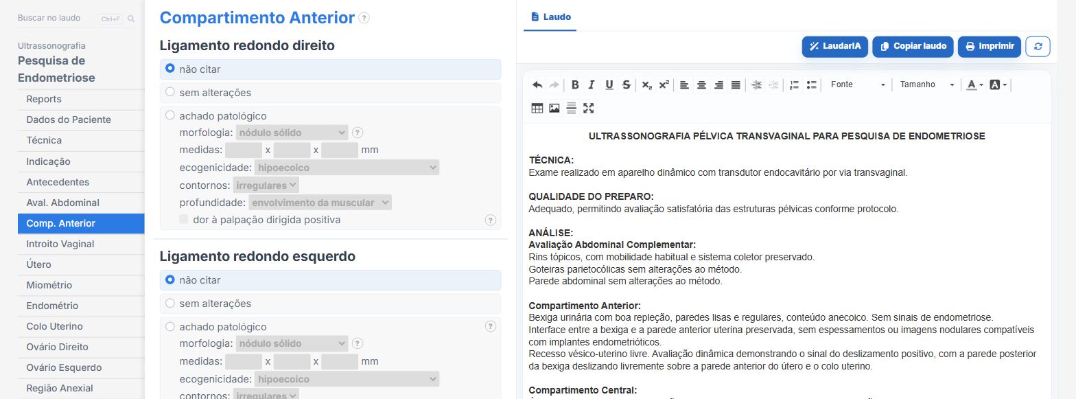
Laudário has a dedicated module for endometriosis assessment, built on the compartment-based taxonomy and designed so that no step of the mapping is forgotten in the rush of routine. In practice, this means:
- Tabs organized exactly in the sequence of the sweep (technique and preparation, complementary abdominal assessment, anterior, central, posterior and lateral compartments, and dynamic assessment), so that the structure of the form guides the exam.
- Fields dedicated to what is usually forgotten: depth of bladder invasion, extrinsic or intrinsic nature of ureteral involvement, involved segment of the uterosacral ligaments, distance from the anal verge and infiltrated layer in bowel lesions.
- Automatic cross-validation between the distance from the anal verge and the bowel segment chosen, with an alert when the two are not anatomically consistent, plus a check of the circumference and stenosis thresholds.
- Standardized phrases with the classic sonographic signs (comet, Indian headdress, mushroom cap, spiculated plaque, diabolo nodule, kissing ovaries) ready and adjustable to your reporting style.
- A preparation quality block that automatically records the corresponding limitation, preventing an exam without preparation from concluding more than it can.
- Report impression assembled automatically from the compartments involved, keeping adenomyosis on a separate line and bringing hydronephrosis, abdominal wall and diaphragm as complementary lines.
- A cartographic map with marking tools for deep endometriosis, endometriomas, adhesions, follicles and adenomyosis, in frontal and sagittal views, printed as a document attached to the report, which turns the exam into a visual roadmap for the surgical team.
- Support from LaudarIA, the integrated artificial intelligence, to help write complex descriptions, such as multicompartmental lesions or patients in postoperative follow-up.
The result is a report that answers the questions the surgical team actually asks, instead of a report that confirms the obvious. In a disease that takes years to diagnose and whose surgery ranges from a simple procedure to a major multidisciplinary operation, the difference between a generic report and a structured mapping is, literally, the difference between the patient being operated on well or being operated on twice.
If you perform endometriosis mapping and still build the report manually or from old text, it is worth trying: Laudário offers a 15-day free trial, no credit card required, with immediate access to all templates, including the complete endometriosis assessment module, transvaginal pelvic ultrasound and the other protocols of the gynecologic routine.
The ultrasound reporting platform built by sonologists. We write about reports, classifications and standardization from the real routine of the practice. Who writes and how.
Educational content for healthcare professionals. It does not replace clinical judgment or the guidelines adopted by your service. Editorial policy.