Kidney and urinary tract ultrasound: how to structure the report from renal measurements to stone evaluation
Kidney and urinary tract ultrasound is one of the most requested exams in clinical practice, from the initial workup of low back pain to the follow-up of patients with chronic kidney disease, long-standing hypertension, recurrent urinary infection or suspected lithiasis. Although it is considered a routine exam, ...
 LaudárioUltrasound reports in practice
This article is part of the guide Abdominal Ultrasound: A Guide to the Findings That Actually Change Management.
Kidney and urinary tract ultrasound is one of the most requested exams in clinical practice, from the initial workup of low back pain to the follow-up of patients with chronic kidney disease, long-standing hypertension, recurrent urinary infection or suspected lithiasis. Although it is considered a routine exam, it is also one in which the difference between a good report and a mediocre one directly affects management. Poorly standardized measurements, generic descriptions of cysts or stones, and conclusions that do not address the indication end up passing the doubt on to the referring physician. In this article, we propose a practical structure for the renal and urinary tract ultrasound report, focused on measurements, echotexture, pelvicalyceal dilation, lithiasis and correlation with the clinical picture.
When kidney and urinary tract ultrasound is requested
The indication varies considerably between services, but there is a recurring set of situations in which renal ultrasound is the first-choice imaging study:
- Low back pain or flank pain: workup for lithiasis, hydronephrosis or renal inflammatory processes.
- Recurrent urinary infection or pyelonephritis: assessment of predisposing factors such as dilations, lithiasis, bladder abnormalities or structural anomalies.
- Gross or microscopic hematuria: screening for masses, complex cysts, lithiasis or bladder abnormalities.
- Chronic kidney disease: morphologic characterization of the kidneys (size, cortical thickness, echogenicity) with direct impact on the diagnosis of chronicity.
- Arterial hypertension: assessment of renal asymmetry, signs of chronic nephropathy or suspicion of a renal/vascular cause.
- Incidental finding on another exam: characterization and follow-up of cysts, solid nodules or dilations.
- Patients with urologic devices: monitoring of double-J stents, nephrostomy or after procedures such as lithotripsy.
Each of these indications calls for a different emphasis in the report. An exam requested for renal colic needs to detail the presence or absence of stones and the degree of dilation. An exam for chronic kidney disease needs, at a minimum, to offer reliable measurements and a careful description of the corticomedullary relationship. When the report does not address the indication, the requesting physician loses an important part of the clinical information.
Recommended structure for the kidney and urinary tract ultrasound report
A good structure for the exam follows, in practice, the anatomical sequence of the upper and lower urinary tract: it starts with identification and technique, describes each kidney individually, assesses the collecting system and the ureters when visible, examines the bladder and closes with the diagnostic impression correlated with the indication. We suggest the following blocks:
1. Identification and technique
Include the type of exam (transabdominal, with or without pre- and post-void bladder assessment), the reason for the request and any technical limitation observed: adiposity, gas-filled bowel loops, difficult positioning, low bladder filling or a poorly cooperative patient. This care puts any limitations in context for the later description and protects the professional in cases of a partial exam.
2. Right and left kidney: measurements and morphology
Describe each kidney in a symmetric, standardized way. Report the longitudinal dimension (ideally also the transverse and anteroposterior dimensions), parenchymal or cortical thickness, position (normal or ectopic), contours (regular, fetal lobulation, irregular due to scarring), cortical echogenicity relative to the liver/spleen and the corticomedullary relationship. In follow-up exams, restating the previous measurements makes it much easier for the referring physician to read the report.
For most adults, consider a longitudinal dimension between 9 and 12 cm and preserved cortical thickness as a reference. Variations outside this range should be interpreted together with the clinical picture: small kidneys with a thinned cortex and increased echogenicity suggest chronicity; symmetrically enlarged kidneys may correspond to infiltrative processes, and asymmetric kidneys call for additional analysis of vascularization and parenchyma.
3. Collecting system and ureters
Describe the presence or absence of pelvicalyceal dilation, grading it when present (mild, moderate, severe). In cases of hydronephrosis, it is useful to mention whether there is an effect on the parenchyma, signs of cortical thinning and, when possible, the level of obstruction. Stones in the collecting system should be localized (upper, middle or lower calyx, pelvis), with approximate size and the presence of posterior acoustic shadowing. When the ureters are not dilated, they are generally not individualized on ultrasound; this should be stated in the report to avoid a mistaken interpretation of “absence” of the structure.
4. Bladder
Even in reports focused on the kidneys, bladder assessment is part of the exam. Describe the contents (anechoic, with debris, with sediment), the wall (thickness, regularity, presence of trabeculation or diverticula) and the presence of vegetating nodular or stone-like images inside. When clinically indicated, record the post-void residual using the ellipsoid formula, especially in patients with lower urinary tract symptoms or suspected bladder outlet obstruction.
5. Focal findings: cysts, nodules and calcifications
Renal cysts should be described with location, size, contours, contents and wall. Simple cysts (anechoic, thin wall, no septations or solid component, with posterior acoustic enhancement) can be categorized as Bosniak I in correlation with CT/MRI, but the ultrasound report needs, at a minimum, to describe these parameters objectively so that the requesting physician knows when the finding is trivial and when it is a complex lesion. Solid nodules, lesions with thickened septations, irregular walls or areas with an apparent solid component deserve emphasis in the conclusion and usually a recommendation for a complementary method.
6. Diagnostic impression
The conclusion should be objective and directly correlate the findings described with the clinical indication. In reports for renal colic, mention the presence/absence of lithiasis, location and associated dilation. In patients with chronic kidney disease, restate the dimensions, cortical thickness and corticomedullary relationship. In patients with hypertension, comment on any asymmetries and signs of chronic nephropathy. The final decision is always clinical: the report only needs to give the requesting physician the elements to make it safely, without assumptions beyond what the method allows.
Common mistakes in the kidney and urinary tract report
Even among experienced professionals, some recurring oversights undermine the quality of the renal ultrasound report:
- Incomplete measurements or in a single plane: reporting only the longitudinal dimension without cortical thickness or without comparison between sides makes follow-up of chronic kidney disease difficult.
- Poorly graded hydronephrosis: using terms such as “slight ectasia” without criteria or alternating between “mild”, “slight” and “minimal” creates noise in comparisons.
- Lithiasis described generically: “hyperechoic images with shadowing” without location, approximate number or size greatly reduces the usefulness of the report for the urologist.
- Cysts without a structured description: lack of information on contents, wall and septations prevents categorization of the cyst and raises doubt about the need for further workup.
- No bladder assessment: in patients with urinary symptoms, omitting the wall, contents and post-void residual impoverishes the exam.
- Conclusion disconnected from the indication: reports requested for renal colic that do not comment objectively on the presence or absence of lithiasis and dilation lose much of their clinical usefulness.
- Poor comparisons: citing a previous exam without restating kidney dimensions, the presence of cysts and stones does not allow the patient's evolution to be followed.
How Laudário helps structure kidney and urinary tract reports
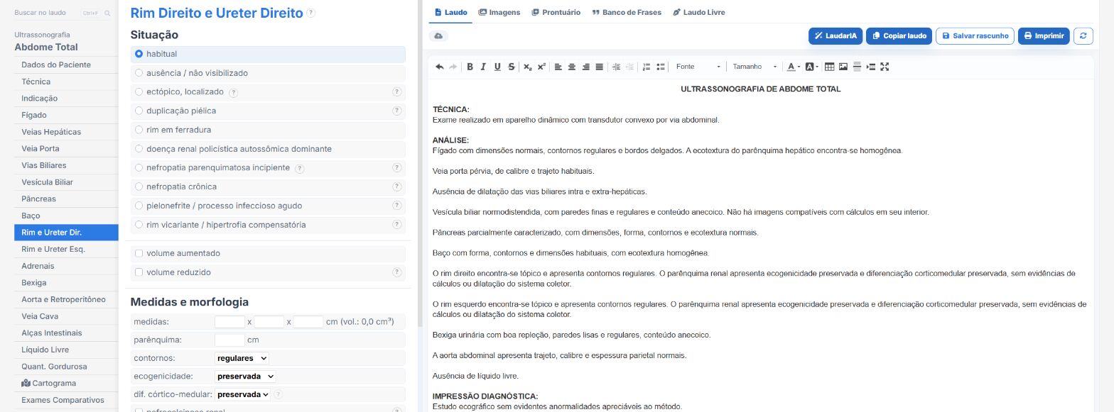
Laudário was designed for professionals who face this routine every day. For kidney and urinary tract ultrasound, and for the main abdominal and urologic protocols, the system offers:
- Ready-made, structured templates for kidneys and urinary tract, total abdomen, prostate via the abdominal route, bladder and pediatric exams, organized in the anatomical sequence of the exam to speed up data entry.
- Standardized, customizable phrases for describing renal morphology, grading hydronephrosis, describing lithiasis and characterizing cysts, keeping terminology consistent across reports.
- Automatic calculations integrated into the data entry flow, including bladder volume and post-void residual by the ellipsoid formula, avoiding manual math and transcription errors.
- Easier comparisons, with automatic retrieval of previous measurements and findings for the same patient, essential for follow-up of chronic kidney disease, cysts under surveillance or recurrent lithiasis.
- A complete library covering kidneys, urinary tract, total abdomen, prostate, thyroid, breast, obstetrics and Doppler, all in a single online system.
The result is simple: less time typing, fewer forgotten findings and more consistent reports across exams of the same patient, which makes all the difference in following patients with chronic kidney disease, recurrent lithiasis or cysts under surveillance. Instead of starting from a blank page for every patient, you adjust an already tested template, keep the clinic's professional standard and focus on what really requires your attention: the careful reading of the images.
The ultrasound reporting platform built by sonologists. We write about reports, classifications and standardization from the real routine of the practice. Who writes and how.
Educational content for healthcare professionals. It does not replace clinical judgment or the guidelines adopted by your service. Editorial policy.