Liver fat quantification by ultrasound: ATI, UGAP, UDFF and how to report
How ultrasound liver fat quantification techniques work, the acquisition protocol that makes the number reliable, why cutoffs vary by vendor and what the report needs to record.
 LaudárioUltrasound reports in practice
This article is part of the guide Abdominal Ultrasound: A Guide to the Findings That Actually Change Management.
Grading steatosis on B-mode (mild, moderate, severe) is subjective and varies between examiners. Ultrasound fat quantification techniques replace this impression with a number, measured reproducibly and comparable between exams. But the number is only valid if the acquisition follows a protocol and the report states how it was obtained.
What each technique measures
Fat in the liver increases the attenuation of the acoustic beam and alters backscatter. Each manufacturer implemented this in its own way, with its own name and unit:
- Canon - ATI (Attenuation Imaging): attenuation coefficient in dB/cm/MHz.
- GE - UGAP (Ultrasound-Guided Attenuation Parameter): attenuation in dB/cm/MHz.
- Siemens - UDFF (Ultrasound-Derived Fat Fraction): estimate of the fat fraction as a percentage, combining attenuation and backscatter.
- Samsung - TAI and TSI (Tissue Attenuation and Tissue Scatter-distribution Imaging), which can be combined into a proprietary index.
- Philips and others: attenuation parameters with different commercial names.
All were validated against MRI proton density fat fraction (MRI-PDFF), which is the noninvasive reference. On MRI-PDFF, values from about 5% indicate steatosis.
Why cutoffs are not interchangeable
An ATI value cannot be compared with a UGAP value, and neither can be compared with the FibroScan CAP, even when the unit is the same. The algorithms are different and so are the published cutoffs. For this reason the report must cite the technology, the value with its unit and the cutoff used, with the reference. And patient follow-up should be done, whenever possible, on the same equipment.
The protocol that makes the number reliable
- Fasting for a few hours: the postprandial state can alter the values.
- Patient in the supine position, right arm raised, intercostal access to the right lobe.
- Region of interest placed about 2 cm from the liver capsule, away from vessels, ducts and reverberation artifacts.
- Measurements during a brief breath hold, at neutral respiration.
- In general, 5 valid measurements, with the result expressed as the median.
- Respect each manufacturer's quality index (coefficient of determination, dispersion or equivalent). A measurement below the quality threshold should not enter the calculation.
Factors that interfere
- Marked obesity and a poor acoustic window.
- Advanced fibrosis, inflammation and storage diseases can interfere with attenuation.
- Focal lesion in the acquisition region.
- Large-volume ascites.
When present, they should be cited in the report, because they change confidence in the number.
What the report should include
- Manufacturer, technology and, if possible, equipment model.
- Acquisition conditions: adequate or limited window, number of valid measurements, lobe assessed, distance from the capsule and confirmation of fasting.
- Value (median) with the unit and the quality index.
- Grading according to the manufacturer's table, with the reference.
- Comparison with prior exams, when available, preferably in an evolution table.
Common errors
- Reporting the value without the technology and the unit.
- Using one manufacturer's cutoff for another manufacturer's equipment.
- Calculating the median including measurements with quality below the threshold.
- Not mentioning that fasting was not confirmed.
- Comparing exams done on different equipment as if they were the same measurement.
See also our articles on hepatic steatosis on B-mode and on liver elastography.
How Laudário helps
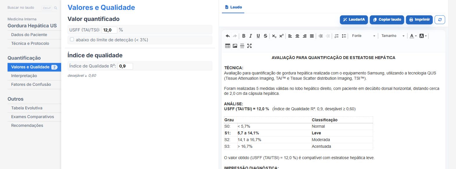
In the liver fat quantification template of Laudário, which can also be included in abdominal reports:
- Technology by manufacturer (Samsung, GE, Canon, Siemens, Philips), with the technique phrase ready.
- Number of measurements, lobe assessed and distance from the capsule in dedicated fields.
- Value and quality index, with a warning when quality falls below the threshold.
- Automatic classification and an evolution table across exams.
Try it free for 15 days, no credit card required.
The ultrasound reporting platform built by sonologists. We write about reports, classifications and standardization from the real routine of the practice. Who writes and how.
Educational content for healthcare professionals. It does not replace clinical judgment or the guidelines adopted by your service. Editorial policy.