Liver ultrasound and hepatic steatosis: how to describe, grade and standardize the report
Hepatic steatosis has gone from an incidental finding to one of the most frequent abnormalities in routine abdominal ultrasound. With the marked rise of nonalcoholic fatty liver disease (NAFLD) in the adult population, and increasingly in adolescents, liver ultrasound...
 LaudárioUltrasound reports in practice
This article is part of the guide Abdominal Ultrasound: A Guide to the Findings That Actually Change Management.
Hepatic steatosis has gone from an incidental finding to one of the most frequent abnormalities in routine abdominal ultrasound. With the marked rise of nonalcoholic fatty liver disease (NAFLD) in the adult population, and increasingly in adolescents, liver ultrasound has become a population screening tool. For exactly that reason, how the finding is described and graded in the report has a direct impact on clinical follow-up, on the ordering of additional tests and on the decision for elastography or biopsy.
Why standardize the description of hepatic steatosis in the report?
Ultrasound is the imaging study most used to screen for hepatic steatosis, yet it is also the study in which variability between reports is most noticeable to the attending physician. It is not uncommon for the same patient to receive very different descriptions in different months: “mild steatosis,” “liver with a diffusely hyperechoic appearance,” “moderate fatty infiltration,” with no clear criteria justifying each category.
The main problems that a lack of standardization causes in this study are:
- Inconsistent grading: what one sonographer calls “mild steatosis” another describes as “moderate,” even on similar images.
- Diagnostic criteria not cited: the report concludes steatosis without mentioning the sonographic signs that support the diagnosis (parenchymal echogenicity, posterior attenuation, visualization of the vasculature).
- No comparison with prior studies: in a chronic disease that evolves over years, comparing the current grade with the previous one is an essential part of follow-up.
- Impression disconnected from the body of the report: the description suggests a mild finding, but the conclusion speaks of severe steatosis, or vice versa.
- No clinical suggestion: the report describes the abnormality but does not guide the next step (follow-up, metabolic profile, elastography).
Recommended structure for the liver ultrasound report
A good structure for liver ultrasound follows the logic of the study: evaluate the gland as a whole, describe focal findings when present, grade the steatosis if there is any, and close with an impression directly correlated with the text. We suggest the following blocks:
1. Identification and technique
Include the type of study, fasting conditions, any technical limitations (body habitus, gas distention, reduced acoustic window) and the equipment, when relevant. This block gives context to the requesting physician and protects the sonographer in technically limited studies.
2. Hepatic parenchyma
Describe dimensions (right and left lobes), contours, echotexture and echogenicity of the parenchyma, comparing with the right renal cortex, the classic reference for evaluating steatosis. Also mention the visualization of the intrahepatic vascular structures and of the diaphragm, since these elements support the grading of the finding.
3. Gallbladder and bile ducts
Even in a study focused on the liver, the gallbladder and bile ducts are a natural part of the evaluation. Describe walls, contents (presence of stones, biliary sludge, polyps), distention and caliber of the intra- and extrahepatic bile ducts. These findings often make up the metabolic context of steatosis.
4. Focal findings
Simple cysts, hemangiomas, areas of periportal or perivesicular focal fatty sparing and solid nodules should each receive their own paragraph, with location (Couinaud segment, when possible), dimensions in three planes, echogenicity, contours and relationship to vessels and ducts. Standardizing this description is what allows future studies to be compared safely.
5. Grading of steatosis
When steatosis is present, it is essential to grade it on objective criteria. The semiquantitative standard most used in practice follows three categories:
- Mild steatosis (grade 1): slight, diffuse increase in hepatic echogenicity relative to the renal cortex, with good visualization of the diaphragm, intrahepatic vasculature and portal branches.
- Moderate steatosis (grade 2): more evident increase in echogenicity, with partial impairment of visualization of the diaphragm and intrahepatic vasculature, and onset of sound beam attenuation in the deeper portions.
- Severe steatosis (grade 3): marked parenchymal hyperechogenicity, significant sound beam attenuation, and difficulty or inability to visualize the diaphragm, intrahepatic vasculature and posterior segments of the right lobe.
Keeping these criteria always in the same terms, and citing them in the body of the report, is what gives consistency to follow-up. In patients with risk factors (metabolic syndrome, diabetes, obesity), it is worth explicitly mentioning these clinical data when they are provided on the requisition.
6. Diagnostic impression and suggested management
The conclusion should be objective, hierarchical and directly correlated with the descriptive body. For steatosis, state the current grade and, when applicable, suggest correlation with metabolic and clinical evaluation, or an additional study with liver elastography for fibrosis stratification, especially in patients with risk factors. The final decision always rests with the attending physician, but the report must provide all the elements to make it.
Common errors in the liver and steatosis report
Even with a defined structure, some recurring errors undermine the quality of liver reports:
- Concluding steatosis without describing the criteria: citing only the grade in the conclusion, while the parenchyma paragraph lacks echogenicity compared with the kidney, posterior attenuation and visualization of the vasculature.
- Underestimating or overestimating the grade: classifying as “mild” a liver with evident posterior attenuation, or as “moderate” a subtle finding, creates clinical noise and makes follow-up harder.
- Forgetting focal fatty sparing: describing a focal hypoechoic area as a new finding, without recognizing the typical periportal or perivesicular pattern, can lead to unnecessary workups.
- Not comparing with prior studies: in a chronic and dynamic disease, failing to cite the previous study means losing the most useful part of follow-up.
- Mixing terminologies: alternating between “steatosis,” “fatty infiltration” and “fatty liver” within the same service, or between “mild/moderate/severe” and “grade 1/2/3,” makes comparative reading harder.
- Conclusion disconnected from the description: speaking of “moderate steatosis” in the impression when the body of the report describes only a slight increase in echogenicity, or vice versa.
How Laudário helps structure liver reports
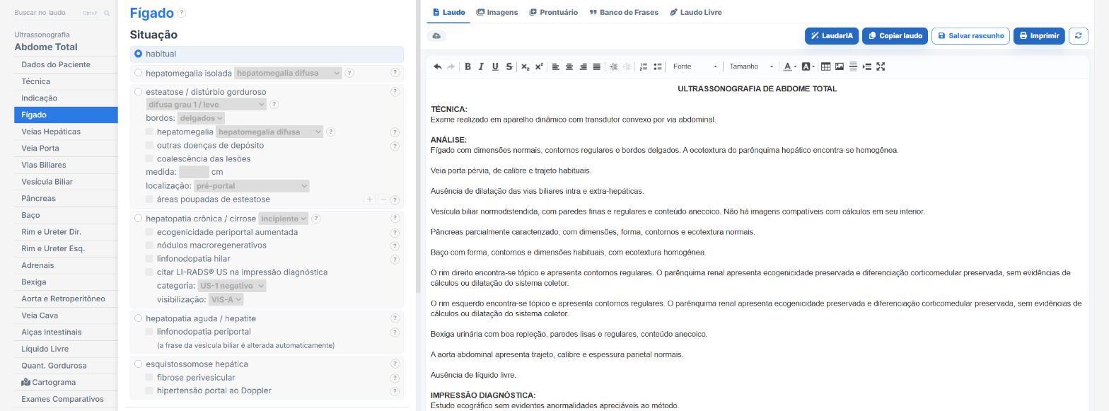
Laudário was designed for professionals who face this routine every day. For liver ultrasound, and for the main abdominal protocols, the system offers:
- Ready-made, structured templates for complete abdomen, upper abdomen and a liver-focused study, organized in the classic sequence of the study to speed up typing.
- Standardized, customizable phrases for describing the hepatic parenchyma, grading steatosis, focal fatty sparing and focal findings, keeping terminology consistent across reports.
- Integrated calculations and measurements in the typing flow, avoiding manual math and reducing transcription errors of dimensions and segments.
- A complete library covering abdomen, pelvis, small parts, obstetrics and Doppler studies, all in a single online system, which also makes standardization among professionals in the same clinic easier.
The result is simple: less time typing, fewer forgotten findings and more consistent reports across studies of the same patient, which makes all the difference in a chronic condition such as steatosis, where sonographic follow-up is central to management. Instead of starting from a blank page for every patient, you adjust an already tested template, maintain the clinic's professional standard and focus on what really demands your attention: reading the images.
If you still assemble your liver reports manually or by copying old text, it is worth getting to know Laudário. Try it free for 15 days and see in practice how a well-designed structure saves time in every study.
The ultrasound reporting platform built by sonologists. We write about reports, classifications and standardization from the real routine of the practice. Who writes and how.
Educational content for healthcare professionals. It does not replace clinical judgment or the guidelines adopted by your service. Editorial policy.