Abdominal Ultrasound: A Guide to the Findings That Actually Change Management
The most requested exam in routine practice is also the most underused. See the abdomen organized by the findings that drive management, from the liver quantification that replaced "mild, moderate, marked" to the aneurysm screening the exam could already be doing.
 LaudárioUltrasound reports in practice
Abdominal ultrasound is the most requested exam in routine practice and probably the most underused. Not because of technical difficulty (the abdomen is where most of us learned to report) but because familiarity produces an automatic report: "liver of normal dimensions, gallbladder without stones, kidneys in normal position". A document that confirms the obvious and rarely decides anything.
This guide organizes the abdomen by the findings that actually change management, organ by organ, and points out where the exam has become something else in recent years, especially in the liver, where quantification has replaced the adjective. Each topic has a dedicated article, linked throughout the text.
Liver: where "mild, moderate, marked" has its days numbered
Liver assessment is the area that has changed the most. For decades, steatosis was graded by visual impression: comparison with the renal cortex, beam attenuation, visibility of vascular walls and of the diaphragm. It is a subjective scale, with modest interobserver agreement, and it is not suited to follow-up: saying that steatosis "improved from moderate to mild" is almost an opinion.
Two technologies changed this, and both are already in routine use:
- Attenuation-based fat quantification (dB/cm/MHz) or fat fraction (%), which gives a number that is comparable between exams. With an important caveat: cutoffs vary by manufacturer. Each technology has its own thresholds for S0 to S3, and applying one device's cutoff to another's value is a silent and common error.
- Elastography, which measures stiffness and answers the question that steatosis does not: does this liver have advanced fibrosis?
And there is an interaction between the two that almost nobody mentions, because it is counterintuitive: they confound each other. Marked steatosis attenuates the ultrasound and degrades elastography; advanced fibrosis overestimates attenuation and inflates the fat estimate. Each method interferes with the other, which is precisely the argument for multiparametric assessment, reading stiffness, fat and B-mode together.
The basics of description and grading are in Liver ultrasound and hepatic steatosis. The deeper dive into stiffness (the SRU consensus Rule of 4, the IQR/M quality criterion, the confounders that overestimate the value and why follow-up is done by the delta and not the absolute value) is in Liver elastography: from the Rule of 4 to confounding factors.
If you take away only one idea from this section: elastography measures stiffness, not fibrosis. Inflammation, congestion and cholestasis make the liver stiff without a single extra fiber. A report that delivers the kPa without the context can induce an error that biopsy would not make.
And there is a third liver exam that completes the set: hepatic Doppler. While elastography measures parenchymal stiffness, Doppler shows its hemodynamic consequence: slow or reversed portal flow, and the collaterals that betray clinically significant portal hypertension. The two exams point to the same outcome by different paths, and corroborate each other.
Gallbladder and bile ducts: the exam that looks simple
Cholelithiasis is the most common abdominal finding and the easiest to report poorly, because the temptation is to report only "presence of stones". The data that change surgical management are others:
- Size of the largest stone and whether it is mobile or impacted, especially in the infundibulum.
- Wall thickness, measured on the anterior wall (the posterior wall undergoes acoustic enhancement and misleads).
- Common bile duct caliber, the datum that separates an elective cholecystectomy from a workup for choledocholithiasis. Remember that it increases physiologically with age and after cholecystectomy.
- Sonographic Murphy sign, the only dynamic finding of the exam, and the one that gives clinical context to the image.
There is also the distinction that generates the most confusion: a polyp does not shadow and does not move; an adherent stone shadows and may not move. The criterion is shadowing and Doppler, not mobility alone. Details in Gallbladder ultrasound and cholelithiasis.
Kidneys and urinary tract: measure and grade
A useful renal report brings three things that the automatic one omits: the measurement of each kidney, the parenchymal thickness (which signals chronic disease before size falls) and, when there is dilation, the grading of hydronephrosis: mild, restricted to the pelvis and major calyces; moderate, with dilation of the fornices; marked, with parenchymal thinning.
Hydronephrosis deserves emphasis because it is the finding that most silently compromises the patient: it can progress to functional loss without any symptom. And it ties into other exams: it is mandatory when ureteral involvement by deep endometriosis is suspected, and it is the marker of upper tract repercussion in obstruction due to prostatic hyperplasia.
The full article is at Kidney and urinary tract ultrasound.
Aorta: the screening the abdominal report could already be doing
This is probably the biggest waste in abdominal routine. The aorta is in the field of view of practically every complete abdominal ultrasound, and abdominal aortic aneurysm is a condition with established cost-effective screening, asymptomatic until it ruptures, and with very high lethality on rupture.
Measuring the maximum anteroposterior diameter, outer wall to outer wall, takes seconds. A man over 65 with a smoking history, having an abdominal ultrasound for another complaint, is exactly the target population for screening, and the exam is already being done. Details in Abdominal aortic ultrasound: aneurysm screening.
Abdominal wall: what is not viscera
The abdomen closes with the wall findings, which tend to fall outside the mental protocol: abdominal wall hernias, whose assessment depends on a dynamic maneuver, with Valsalva and in the upright position, because a reducible hernia simply does not appear at rest, and soft tissue lesions. A painful, cyclic nodule in a cesarean scar, for example, has an explanation that is rarely considered: abdominal wall endometriosis.
The errors that most often appear in the abdominal report
- Grading steatosis without explicit criteria and calling the variation between exams improvement or worsening.
- Reporting kPa without the IQR/M: without it, the clinician cannot know whether the number is reliable.
- Applying one manufacturer's fat cutoffs to another's value.
- Comparing elastographies from different devices: variability between systems equals the threshold of clinical significance, which nullifies the comparison.
- Measuring the gallbladder wall on the posterior side, overestimated by acoustic enhancement.
- Not measuring the common bile duct when there is cholelithiasis.
- Not grading hydronephrosis: "pelvicalyceal dilation" does not indicate management.
- Not measuring the aorta in a patient from the screening population, with the exam already under way.
- Not measuring the kidneys and concluding "normal dimensions".
How Laudário helps
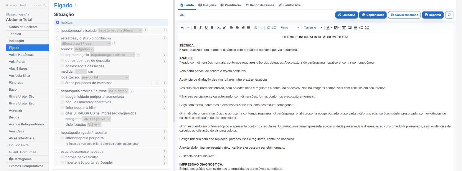
Laudário covers the entire abdomen: upper and complete abdomen, with and without prostate, urinary tract, hepatic and aortic Doppler, liver elastography, fat quantification and multiparametric assessment. In practice:
- Steatosis cutoffs by manufacturer technology, applied automatically according to the device, with no risk of using the wrong threshold.
- Automatic elastography classification by the Rule of 4, with IQR/M calculated, a caveat for non-validated etiology and the follow-up delta against the 10% threshold.
- Automatic volume calculation and an alert when a measurement falls outside the reference range.
- Evolution table that cross-references stiffness, fat fraction and date in the same grid.
- Dedicated fields for what tends to be omitted: common bile duct, renal parenchymal thickness, aortic diameter.
- Support from LaudarIA for complex interpretations: multiple confounders, long follow-ups, findings that contradict each other.
If your abdominal report is still copied from an old text, it is worth trying: 15 days free trial, no credit card, with all templates unlocked.
The ultrasound reporting platform built by sonologists. We write about reports, classifications and standardization from the real routine of the practice. Who writes and how.
Educational content for healthcare professionals. It does not replace clinical judgment or the guidelines adopted by your service. Editorial policy.