Prostate ultrasound: how to structure the report for prostate volume, BPH and nodules
A useful prostate report gives the three measurements, the volume by the ellipsoid formula (D1 x D2 x D3 x 0.52) with the enlargement range, the median lobe and nodule description, and the post-void residual with its clinical meaning.
 LaudárioUltrasound reports in practice
A prostate ultrasound report that is truly useful to the urologist needs to deliver five pieces of information objectively: the three dimensions of the gland in millimeters, the prostate volume calculated with the simplified ellipsoid formula (D1 × D2 × D3 × 0.52) in cubic centimeters, the characterization of the contour, echotexture and any nodules, the presence and intravesical protrusion of the median lobe in millimeters, and the post-void residual measured under adequate conditions. Volume and residual are what guide management of symptomatic benign prostatic hyperplasia (BPH), and the description of focal findings is where the first suspicion arises that directs PSA, multiparametric MRI and, eventually, biopsy.
The reference ranges widely used in urologic practice guide the conclusion: up to 30 cm³ the volume is considered normal; between 30 and 40 cm³ there is mild enlargement; between 40 and 80 cm³, moderate enlargement; above 80 cm³, marked enlargement, with a direct impact on the choice of surgical technique. For post-void residual, values up to 50 ml are normal, between 50 and 100 ml call for clinical attention, and above 100 ml suggest significant bladder outlet obstruction. Standardizing these numbers and how they are obtained is what separates a report that drives management from one that merely confirms an “enlarged prostate”.
Why standardize the prostate report
The prostate is the male organ in which ultrasound most often makes clinical decisions “on its own”: prostate volume and post-void residual are the two numbers the urologist looks at first to define management of symptomatic BPH, whether to start medical treatment or to indicate surgical relief of obstruction. When these values reach the chart with different technical criteria at each study, follow-up loses meaning: a “prostate that grew” may simply reflect a change in calculation method, not in disease.
Without standardization, the same patient receives reports with volumes that vary by 10 to 20% just because of how they were measured, with inconsistent description of the median lobe and intravesical growth, and with post-void residuals measured without attention to the pre-void bladder volume. A standardized report describes the dimensions on all three axes, calculates the volume with the ellipsoid formula, characterizes the contour, echotexture and the presence of nodules, measures the median lobe when prominent, assesses the post-void residual under adequate conditions, and concludes by explicitly stating the clinical meaning of the findings.
Indications and data that should appear in the identification
The report should state the reason for the study right at the start, because it completely changes what will be emphasized in the description and conclusion. The most common indications include:
- Lower urinary tract symptoms (LUTS), both storage and voiding: weak stream, hesitancy, terminal dribbling, nocturia and urgency. Here the report needs to detail the impact on the bladder and the residual.
- Elevated or monitored PSA, either to correlate prostate volume with PSA density or to guide the decision on follow-up or biopsy. A study for elevated PSA must, at the very least, provide a reliable volume for the density calculation.
- Follow-up of known BPH and post-treatment monitoring, medical or surgical.
- Suspected chronic urinary retention, with the post-void residual as an indirect functional measure of obstruction.
- Hematuria, recurrent urinary tract infections in men, and prostatitis, often complementary to the evaluation of the upper urinary tract.
When the study is serial, record the date of the prior study and state explicitly whether the comparison shows stability, volumetric progression or reduction after medical treatment: 5-alpha-reductase inhibitors, for example, can reduce volume by 20 to 30%. In patients with previous surgery (TURP, simple prostatectomy, HoLEP, adenomectomy, radical prostatectomy), the indication should state this clearly, because the anatomic interpretation changes completely. When the report does not address the indication, the referring physician loses an important part of the clinical information.
Zonal anatomy: what each zone means in the report
The prostate is a retroperitoneal pelvic gland, located inferior to the bladder, anterior to the rectum, and traversed by the prostatic urethra. For the report, it is useful to think of three zones with distinct clinical implications:
- The peripheral zone is the largest glandular portion and is the site of origin of about 70% of prostate adenocarcinomas. On ultrasound it usually appears as the posterior third of the parenchyma, with slightly more homogeneous echotexture.
- The transition zone surrounds the prostatic urethra and is the characteristic site of BPH: when it enlarges, it accounts for the volumetric growth seen on the study.
- The central zone, around the ejaculatory ducts, is rarely a primary source of pathology, but it is where utricle cysts and dilatations associated with infertility are located.
The two approaches and what each allows you to describe
The choice, or combination, of acquisition approaches completely changes what can be described, and this must be explicit already in the technique block.
Abdominal (suprapubic) approach
This is the standard approach for prostate volume, effect on the bladder, bladder wall and post-void residual, and the approach of choice for screening of BPH and LUTS in most outpatient services. It requires a moderately full bladder (ideally 200 to 300 ml), with a 3.5 to 5 MHz convex transducer, in transverse and longitudinal planes. The three basic measurements are the transverse diameter (width, on the largest transverse section), the anteroposterior diameter (thickness, on the same transverse section) and the longitudinal diameter (height, on the midsagittal section). Its natural limitations are the low resolution for the internal zones of the gland, dependence on bladder filling, and interference from obesity or abdominal scars.
Transrectal approach (TRUS)
With a high-frequency endocavitary transducer, it offers far superior resolution for zonal anatomy, characterization of focal nodules, prostatic capsule, seminal vesicles, Doppler study and targeted biopsy. It is the preferred approach when there is clinical suspicion of neoplasia, detailed investigation of the peripheral zone, chronic pelvic pain, hematospermia, infertility, suspected prostatitis or biopsy planning, and the most suitable for detailing focal findings in patients with elevated PSA.
In combined reports, describe the suprapubic evaluation first, with volume and bladder, and then the transrectal evaluation, with zonal detail, making clear which finding was better characterized by each approach. This avoids confusion and gives the urologist a more useful document.
Diagnostic criteria: how to interpret the volume
Prostate volume is a widely used reference in urologic practice for classifying glandular growth and guiding treatment:
- Normal-volume prostate: up to 30 cm³. It does not exclude disease, but it rules out volumetrically significant BPH as the cause of obstructive symptoms.
- Mild enlargement: 30 to 40 cm³. Frequently consistent with early symptomatic BPH, with indication for medical treatment in symptomatic patients.
- Moderate enlargement: 40 to 80 cm³. Typical range of BPH with an established indication for medical treatment and, in refractory cases, surgical relief of obstruction.
- Marked enlargement: above 80 cm³. Considered a marker of a “large prostate”, with a direct impact on the choice of surgical technique (HoLEP, open or robotic adenomectomy, rather than conventional TURP).
These cutoffs should appear, even if briefly, in the conclusion of the report. The sonologist does not replace the urologist, but a report that merely records “prostate of 72 cm³” without commenting on the enlargement range and clinical meaning delivers less value than it could.
How to describe the findings in the report
Once volumetric enlargement or a focal finding is identified, the description needs to be complete enough for the urologist to decide between follow-up, supplementation with PSA, multiparametric MRI or biopsy without having to repeat the study. The minimum elements are:
- Dimensions on the three axes, in millimeters, with the volume calculated by the ellipsoid formula in cubic centimeters.
- Contours: regular or lobulated; symmetry between the lobes; presence and degree of bulging at the bladder base.
- Echotexture: homogeneous or heterogeneous, with description of the distribution.
- Calcifications: presence, location (parenchymal, periurethral, capsular) and dimensions of macrocalcifications; small punctate calcifications are common and rarely clinically relevant.
- Nodules: when present, describe location (peripheral, transition, central zone), dimensions, echogenicity (hypo, iso, hyper), contour and margins, vascularity on color Doppler when available, and relationship with the capsule. Hypoechoic nodules in the peripheral zone deserve emphasis because of their greater association with neoplasia.
- Median lobe: presence, dimensions and degree of intravesical protrusion in millimeters; it is a clinically decisive finding, because patients with a prominent median lobe respond worse to medical treatment alone and frequently progress to an indication for surgery.
- Prostatic capsule: describe continuity and regularity; signs of bulging or interruption may indicate advanced disease and require supplementation with targeted imaging.
- Seminal vesicles: caliber, symmetry and presence of dilatations, cysts or calcifications. Minor asymmetries are common, but marked dilatations or heterogeneous content deserve emphasis, especially in patients with hematospermia or infertility.
Accompanying findings: what must not be missed
The prostate study is rarely requested in isolation: it almost always comes with an evaluation of the lower urinary tract, and some accompanying findings must be actively sought because they directly change management:
- Post-void residual (PVR): a mandatory measurement in any prostate ultrasound for obstructive symptoms. Calculated with the ellipsoid formula in the bladder after spontaneous and complete voiding. Values up to 50 ml are considered normal; between 50 and 100 ml call for clinical attention; above 100 ml suggest significant bladder outlet obstruction and raise the risk of complications such as urinary tract infections, bladder stones and chronic urinary retention. It is worth stating the time between voiding and acquisition of the residual image, since late measurements may underestimate the true residual.
- Bladder wall: diffuse thickening (> 5 mm with a distended bladder) is an indirect marker of chronic obstruction and should be described. Trabeculations, diverticula and cellules suggest a stressed (trabeculated) bladder. A trabeculated bladder indicates significant chronic obstruction even when the prostate volume is not exuberant.
- Bladder content: anechoic or with debris and sediment.
- Bladder stones: common in patients with prolonged obstruction; describe number, location and dimensions.
- Bladder tumors: papillomas, vegetating lesions or focal thickenings should be characterized and deserve emphasis in the conclusion.
- Hydronephrosis: when assessed together, signs of bilateral pelvicalyceal dilatation indicate obstructive repercussion on the upper urinary tract, with immediate clinical implications.
- Inadequate bladder distension: record it when the study is limited by an underfilled bladder, since this compromises both the volume measurement and the evaluation of the wall and median lobe. The same applies to adipose panniculus, scars and intolerance of the transrectal approach.
Conclusion and clinical guidance
The conclusion of the prostate report should synthesize, in a few lines, three essential pieces of information for the requesting physician: the prostate volume in cubic centimeters with the corresponding enlargement range, the presence or absence of suspicious focal findings, and the post-void residual with its clinical meaning. When the study is normal, an objective conclusion (“prostate of preserved dimensions and echotexture, with estimated volume of XX cm³, without evidence of nodules or intravesical growth, and post-void residual within normal limits”) is more valuable than generic descriptions such as “prostate without abnormalities”.
In patients under follow-up, the conclusion must state the comparison with the prior study explicitly: volumetric stability, progression or reduction after medical treatment. Simply repeating the volume without contextualizing the comparison is a common failure that substantially reduces the clinical value of the serial report. The final decision is always clinical: the report must give the urologist the elements to make it safely.
Findings that deserve emphasis in the conclusion
- Prostate volume above 30 cm³, with the corresponding range and suggested clinical correlation.
- Hypoechoic nodules of the peripheral zone, nodules with anomalous vascularity or capsular bulging, with an explicit suggestion of correlation with PSA and specialist evaluation.
- Median lobe with relevant intravesical protrusion, even in prostates of moderate volume.
- Elevated post-void residual (above 100 ml), with a suggestion of urologic evaluation.
- Signs of a stressed bladder (wall thickening, trabeculations, diverticula, bladder stones), markers of chronic obstruction.
- Associated bilateral hydronephrosis, a sign of repercussion on the upper urinary tract, with indication for immediate evaluation.
- Significant technical limitation from inadequate bladder distension or gas interposition, when the study did not allow a reliable measurement: transparency here prevents false negatives.
Common errors that make the report ambiguous
- Volume without a clear method: always calculate with the ellipsoid formula using the three measurements on the correct axes and state the method; reporting an “enlarged prostate” without an objective volume, or using measurements obtained in different planes at each study, undermines follow-up.
- Oblique section: measure the prostate on a truly midsagittal section, because oblique sections overestimate the height and the final volume.
- Inadequate bladder filling: an empty bladder rules out the acoustic window; an overdistended bladder flattens the prostate and underestimates the height.
- Missing or late residual: always measure right after spontaneous voiding, with no new fluids in the interval; in patients who did not void adequately, record the limitation instead of omitting it.
- Median lobe without a number: describe the intravesical protrusion in millimeters, and not just as “prominent”: this detail changes the surgical decision.
- Inconsistent terminology: alternating between “middle lobe”, “median lobe” and “median portion” in the same report creates noise; the same applies to the description of nodules, which should keep the same wording throughout the text.
- Poorly qualified calcifications: differentiate parenchymal calcifications, without significant clinical repercussion, from prostatic stones associated with chronic prostatitis, with adequate clinical correlation.
- Nodule merely recorded: when there is a hypoechoic nodule in the peripheral zone, describe dimensions, location and capsular relationship and explicitly suggest correlation with PSA and multiparametric MRI.
- Poor comparisons: citing prior studies without restating prostate volume, residual and bladder dimensions does not allow the patient's evolution to be followed.
How Laudário helps structure the prostate report
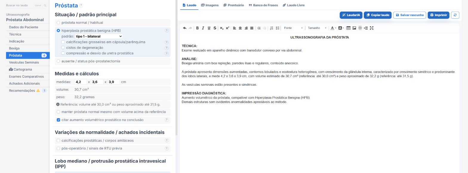
Laudário was developed to speed up report typing without losing the clinical depth that studies such as prostate ultrasound require. The abdominal and transrectal prostate templates, urinary tract and bladder templates come with the structure ready, organized in the order of acquisition. In practice, this means:
- A ready structure to describe each zone of the prostate, with standardized phrases adjustable to your reporting style, keeping terminology consistent between reports.
- Automatic calculation of prostate volume and post-void residual with the ellipsoid formula from the entered measurements, eliminating the risk of arithmetic error during fast typing and standardizing the method across successive studies.
- Dedicated fields for nodules, median lobe, bladder wall and calcifications, so that important criteria are not forgotten.
- A standardized conclusion, adjustable for both normal studies and reports with focal findings, with automatic suggestion of clinical correlation according to the volume range and the presence of suspicious nodules.
- Integration with the urinary tract and bladder templates, facilitating combined reports when the study identifies associated hydronephrosis, bladder wall thickening, diverticula or bladder stones, within a library that also covers total abdomen, thyroid, breast, obstetrics and Doppler.
- Professional printing with a QR Code and a digital version of the report, adding traceability and more perceived value in delivery to the patient and the requesting physician.
- Support from LaudarIA, the integrated artificial intelligence, to help build phrases, conclusions and fine adjustments to the text, particularly useful in complex descriptions such as heterogeneous prostates, multiple nodules or patients in follow-up after medical or surgical treatment.
The result is a standardized prostate report that is quick to produce and, above all, more useful to the urologist who will receive the study. In a high-volume ultrasound practice, standardizing the measurement of prostate volume and keeping the description of the median lobe, the post-void residual and the focal findings always complete means less rework, more diagnostic confidence and greater added value in every study delivered, especially in a condition in which the volume recorded in the report is, literally, part of the equation that defines the surgical indication.
If you still build your prostate reports manually or by copying old texts, it is worth trying: Laudário offers a 15-day free trial, with no credit card, with immediate access to all exam templates, including suprapubic and transrectal prostate, urinary tract and the other protocols of the urologic routine.
The ultrasound reporting platform built by sonologists. We write about reports, classifications and standardization from the real routine of the practice. Who writes and how.
Educational content for healthcare professionals. It does not replace clinical judgment or the guidelines adopted by your service. Editorial policy.