Peyronie disease and penile fracture on ultrasound: plaques, curvature and what to report
How to describe Peyronie disease plaques, what ultrasound says about active and stable phases, when to add pharmacologically induced Doppler and what must not be missing from the report when penile fracture is suspected.
 LaudárioUltrasound reports in practice
Penile ultrasound is the imaging exam of choice both in Peyronie disease and in suspected penile fracture. They are very different situations, one chronic and the other an emergency, but in both the main finding lies in the same structure: the tunica albuginea, the fibrous layer that surrounds the corpora cavernosa.
Technique
- High-frequency linear transducer, with the penis in the anatomical position, resting on the abdomen.
- Sweep in transverse and longitudinal planes, from the base to the glans, over the dorsal, ventral and lateral surfaces.
- Assessment of the corpora cavernosa, corpus spongiosum, urethra and intercavernous septum.
Peyronie disease: the plaques
Plaques appear as focal thickening of the tunica albuginea, usually hyperechoic, with or without acoustic shadowing. For each plaque, the report should state:
- Location: dorsal surface (the most common), ventral, right or left lateral, or septum.
- Third: proximal, middle, distal or junction with the glans.
- Measurements, preferably in all three dimensions, for comparison at follow-up.
- Appearance: hyperechoic without shadowing (fibrotic), with shadowing (calcified), hypoechoic with thickening, or focal loss of continuity.
- Calcification: absent, punctate or extensive with shadowing.
Calcification tends to appear in older plaques and is usually associated with the stable phase, but the phase of the disease is defined clinically (pain, progression of curvature). The report describes; it should not state the phase based on appearance alone.
Curvature and erectile function
Curvature is best assessed in erection. When the urologist needs to plan surgery, the exam with pharmacologic induction allows the angle to be measured and penile Doppler to be added to assess arterial and venous function, which changes the surgical technique indicated. Otherwise, curvature reported by the patient or by photograph can be recorded as clinical data.
Penile fracture: an emergency
Fracture is rupture of the tunica albuginea with the penis erect. Ultrasound should answer quickly:
- Is there a break in the continuity of the tunica albuginea? Location (side, surface and third) and extent of the defect.
- Hematoma: whether it is contained by Buck fascia or extends to the subcutaneous tissue, scrotum or perineum.
- Corpus spongiosum and urethra: an associated injury changes the surgery and requires urologic assessment of the urethra.
Precise localization of the defect helps the surgeon choose the incision. When ultrasound does not show the defect but the clinical picture is typical, the report should state that the finding does not exclude fracture.
Common mistakes
- Describing a "Peyronie plaque" without location, third and measurements.
- Stating the active or stable phase based only on echogenicity.
- In fracture, not describing the corpus spongiosum and urethra.
- Not mentioning the extent of the hematoma.
- A report of "no abnormalities" without stating that the tunica was assessed along its entire length.
See also our text on penile Doppler in erectile dysfunction.
How Laudário helps
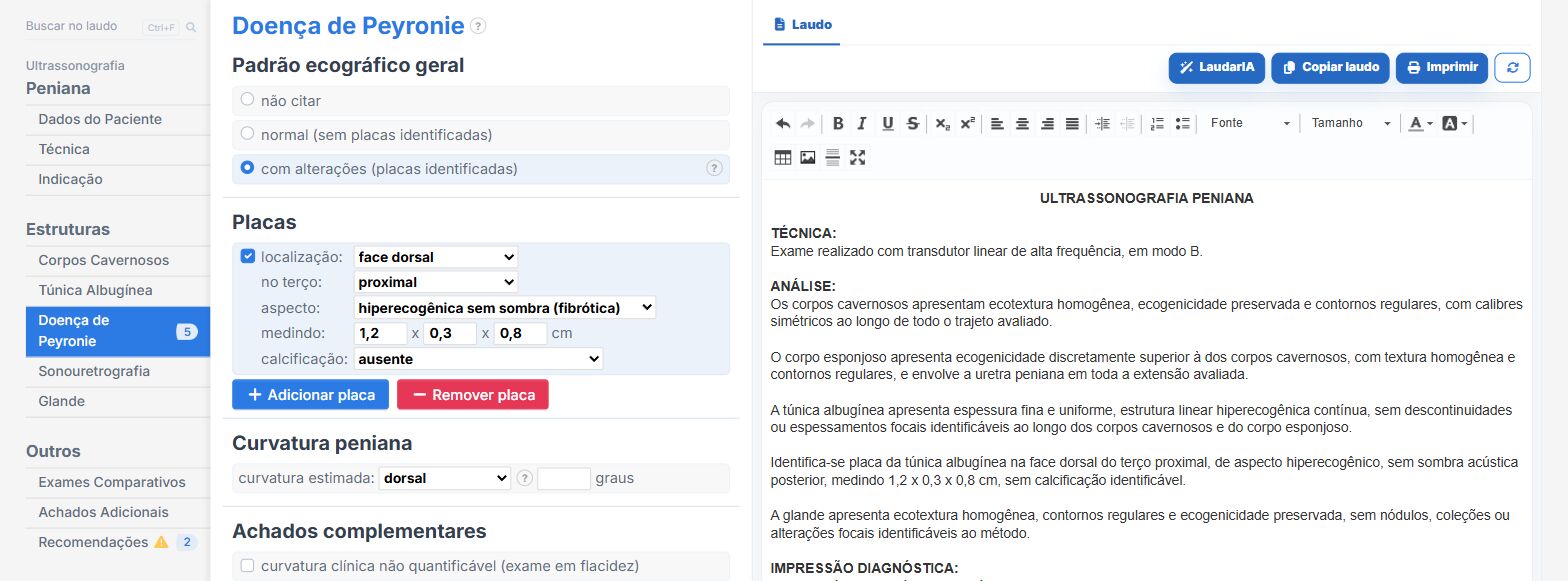
In the penile ultrasound templates of Laudário, with and without Doppler:
- Peyronie plaques with location, third, appearance, measurements and calcification, one entry per plaque.
- Curvature, tunica albuginea, corpora cavernosa, corpus spongiosum and sonourethrography in their own tabs.
- In the Doppler template, pharmacologic induction and velocities in their own fields.
Try it for 15 days free, no credit card required.
The ultrasound reporting platform built by sonologists. We write about reports, classifications and standardization from the real routine of the practice. Who writes and how.
Educational content for healthcare professionals. It does not replace clinical judgment or the guidelines adopted by your service. Editorial policy.