Penile Doppler in erectile dysfunction: the protocol, the velocities and how to report arterial and venous insufficiency
How Doppler is performed after intracavernosal injection, which measurements to record and at what times, peak systolic and end-diastolic velocity values, and what else to describe, such as Peyronie plaques.
 LaudárioUltrasound reports in practice
Penile Doppler is the study that differentiates the vascular causes of erectile dysfunction: arterial insufficiency and veno-occlusive dysfunction. It is a protocol-based study, and interpretation depends on an adequate erection. The report needs to state how the study was done, at what times the measurements were taken and what the response was.
The protocol
- Baseline study in the flaccid state: corpora cavernosa, tunica albuginea, urethra and the cavernosal arteries.
- Intracavernosal injection of a vasoactive drug, generally prostaglandin E1, given by the responsible medical team.
- Measurements in both cavernosal arteries, at the base of the penis, at serial time points, for example at 5, 10, 15 and 20 minutes.
- Recording of the quality of the erection obtained. Anxiety can reduce the response, and a second dose or stimulation may be needed.
Prolonged erection is a known complication of the injection, and the service must have a defined guidance and management plan for it.
The values
- Peak systolic velocity (PSV) of the cavernosal artery: values above about 35 cm/s indicate adequate arterial function; below 25 cm/s indicate arterial insufficiency; between 25 and 35 cm/s, the interpretation is borderline.
- End-diastolic velocity (EDV): with an adequate arterial response, EDV persistently above about 5 cm/s indicates veno-occlusive dysfunction (venous leak).
- Resistive index (RI): values close to 1, with absent or reversed diastolic flow, indicate normal veno-occlusion; lower values reinforce the suspicion of leak.
Veno-occlusive dysfunction can only be diagnosed when the arterial response is adequate. With a low PSV, an elevated EDV cannot be interpreted.
What else to describe
- Peyronie disease plaques: focal thickening of the tunica albuginea, with or without calcification, with location and dimensions.
- Asymmetry between the cavernosal arteries.
- Early reversed flow and collaterals.
- Fibrosis of the corpora cavernosa.
Common errors
- Reporting arterial insufficiency with a clearly incomplete erection, without recording the response.
- Diagnosing venous leak with a low PSV.
- Measuring velocities far from the base of the penis or at an inadequate angle.
- Omitting the time points of the measurements.
See also the articles on scrotal ultrasound with Doppler and prostate ultrasound.
How Laudário helps
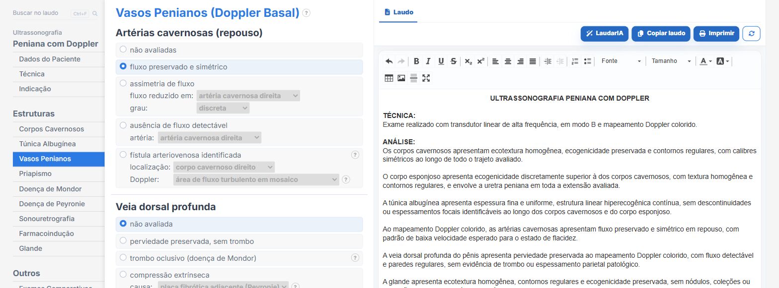
In the penile Doppler templates of Laudário:
- Velocities of both cavernosal arteries by time point, in a table.
- Peyronie plaques with location and measurements.
- Technique, drug and response recorded in the technique tab.
Try it free for 15 days, with no credit card.
The ultrasound reporting platform built by sonologists. We write about reports, classifications and standardization from the real routine of the practice. Who writes and how.
Educational content for healthcare professionals. It does not replace clinical judgment or the guidelines adopted by your service. Editorial policy.