Scrotal ultrasound with Doppler: how to structure the report for pain, varicocele and testicular masses
The scrotal ultrasound report should always follow the same sequence (technique, testes, epididymides, tunicae, pampiniform plexus and Doppler) and end with a clear impression that answers the clinical question: torsion, epididymo-orchitis, varicocele and its grade, or a suspicious mass.
 LaudárioUltrasound reports in practice
Scrotal ultrasound is one of the most sensitive studies for investigating acute testicular pain, scrotal swelling, male infertility and palpable masses. It is a short study, but its clinical weight is out of proportion to the time spent at the scanner: within a few minutes, the sonologist must distinguish inflammatory pain from testicular torsion, describe varicoceles that may affect fertility, assess solid masses with malignant potential, and still deliver a clear impression that guides the urologist or the emergency department.
The practical answer is standardization. A well-structured report always follows the same sequence: technique, right testis, left testis, epididymides, tunicae and scrotal contents, pampiniform plexus and vascular assessment with Doppler. It ends with a prioritized diagnostic impression that unequivocally answers the reason for the request: are there signs of testicular torsion or not, is there epididymo-orchitis, is there varicocele and of what grade, is the mass suspicious or benign. A study like this avoids unnecessary surgical exploration, guides urologic management and protects the physician from a medicolegal standpoint.
Why standardize the scrotal ultrasound report
The scrotum contains small, symmetric structures with a very characteristic sonographic pattern. Small variations in the description (testicular volume, presence or absence of Doppler flow, caliber of the pampiniform plexus veins) have a direct impact on management. A vague report that simply concludes “no abnormalities” leaves gaps in conditions such as microcalcifications, subclinical varicocele, epididymal cysts and small testicular masses, all of which have relevant practical implications.
When a practice adopts a single structure, always covering the same elements, the benefits are clear:
- Greater diagnostic confidence in acute pain, with rapid differentiation between torsion, epididymo-orchitis and trauma.
- Reliable comparison across serial studies (follow-up after epididymo-orchitis, microlithiasis surveillance, after varicocele surgery).
- Standardized description of varicocele, with a grading that is reproducible between examiners.
- Robust documentation in suspicious masses referred for surgical management.
Report structure, block by block
The sequence below follows the logic of the scan and makes quick reading easier for the referring urologist.
1. Identification and technique
State the type of study (with or without color and spectral Doppler), the clinical indication, the equipment when relevant, the patient position, use of the Valsalva maneuver and any technical limitations: a large scrotum, severe pain that limits compression, or a large hydrocele that moves the structures away from the transducer. This block protects the examiner and informs the referring physician about restrictions of the study.
2. Right testis and left testis
Each testis deserves its own paragraph, always with the same sequence of items, so that comparison between sides is immediate:
- Position and contours: usual position in the scrotum, regular or irregular contours.
- Dimensions in three planes and volume: length, width and height, with volume calculated by the ellipsoid formula. Symmetry between the testes is clinically relevant, especially when investigating infertility or atrophy.
- Echotexture and echogenicity: homogeneous or heterogeneous, usual pattern or diffuse hypoechogenicity, presence of testicular microlithiasis (and, in that case, graded by the number of foci per field).
- Focal masses: describe each relevant mass individually, with location (upper, middle or lower third), dimensions in three planes, echogenicity, contours, presence of calcifications or cystic areas, vascularity pattern on color Doppler and relationship with the tunica albuginea.
- Doppler vascularity: presence of intratesticular arterial and venous flow, symmetry between sides, preserved or altered arterial spectral waveform and resistive indices when indicated, especially in acute presentations.
In the setting of acute pain, direct comparison of vascularity between the two testes at the same Doppler settings is essential information and should appear explicitly in the report.
3. Epididymides
Describe the head, body and tail of each epididymis separately, with head dimensions, echotexture, echogenicity and vascularity. Thickening, increased caliber, hyperemia on Doppler and cystic masses (spermatoceles) should be reported with laterality and precise location. Tail thickening in particular is a key finding in epididymo-orchitis, a diagnosis that benefits greatly from objective descriptions compared between the sides.
4. Tunicae, extratesticular contents and scrotal wall
Report the presence and estimated volume of hydrocele, fluid characteristics (anechoic, with debris, septations), thickening of the tunicae and scrotal wall, organized collections (hematocele, pyocele), inguinoscrotal hernias, and cysts of the tunica albuginea or tunica vaginalis. This block prevents frequent findings from appearing generically in the impression.
5. Pampiniform plexus and varicocele assessment
Assessment for varicocele is a chapter of its own in the scrotal report and deserves its own description: laterality, maximum caliber of the pampiniform plexus veins at rest and during the Valsalva maneuver, presence, duration and velocity of venous reflux on Doppler, upright position when indicated, and the classification used by the practice (Sarteschi, Dubin-Amelar or another adopted institutionally). Simply mentioning “plexus veins of increased caliber” leaves the urologist without enough information to decide between watchful waiting and surgery, and standardized phrases prevent the same patient from receiving very different descriptions on successive studies.
6. Diagnostic impression and suggested management
The impression should be objective and prioritized, starting with the findings of greatest clinical relevance. In acute presentations, state unequivocally whether or not there are findings suggesting testicular torsion, epididymo-orchitis or other causes of scrotal pain. In elective studies, highlight varicoceles and their grade, solid masses, microlithiasis, hydrocele and estimated volume, and any other findings that deserve follow-up or clinical correlation. The final decision always rests with the treating physician, but the report should give them every element needed to make it safely.
In emergency studies, this structure becomes even more important. The on-call physician usually reads only the impression before deciding on management. A clear closing statement, with explicit laterality and Doppler comparison between sides, avoids ambiguous interpretations in testicular torsion, where every minute can compromise testicular viability.
Most common clinical scenarios
Acute scrotal pain
Here the report must directly answer the question of testicular torsion versus epididymo-orchitis and other causes (torsion of the appendix, incarcerated hernia, trauma). Color and pulsed Doppler are central: absent or markedly reduced intratesticular arterial flow, compared with the contralateral side, supports the diagnosis of torsion. In contrast, diffuse increased vascularity of the epididymis and/or testis, with a thickened, hypoechoic epididymis, points to an inflammatory/infectious process.
Varicocele and infertility workup
In patients referred for infertility or chronic discomfort, assessment of the pampiniform plexus should be explicit, with caliber at rest and on Valsalva, reflux and grading. Describing only the venous caliber at rest, without reporting behavior during the maneuver, leaves the workup incomplete. Testicular volume on each side is equally central in this indication and in hypogonadism, both in the comparison between sides and over time.
Testicular mass
Every solid testicular mass should be treated as potentially neoplastic until proven otherwise. The report must characterize size, number, location, echogenicity, associated microcalcifications, Doppler vascularity and relationship with the tunica albuginea. Cystic lesions should be differentiated from epididymal cysts, spermatoceles and cysts of the tunica albuginea, with a clear description of the anatomic location.
Common errors in the scrotal ultrasound report
- Concluding “normal study” without reporting the testicular volume on each side and the symmetry.
- Omitting assessment of intratesticular arterial flow in patients with acute pain: presence, symmetry and pattern should appear in the body of the report and, ideally, in the impression as well.
- Describing varicocele without detailing laterality, venous caliber, behavior on Valsalva and the classification adopted.
- Mentioning only “nodular image” or “testicular nodule” without saying in which testis, in which third, with what dimensions, echogenicity, contours and vascularity pattern.
- Failing to differentiate epididymal cyst, spermatocele and cyst of the tunica albuginea, lumping them all together as “cystic images”.
- Poor comparisons: not citing prior studies when they exist, or using vague phrases such as “stable compared with prior” without restating the dimensions of the masses or the varicocele.
- A generic impression, such as “nonspecific changes” or “correlate clinically”, that does not answer the clinical reason for the request (acute pain, infertility, palpable mass).
How Laudário helps with the scrotal ultrasound report
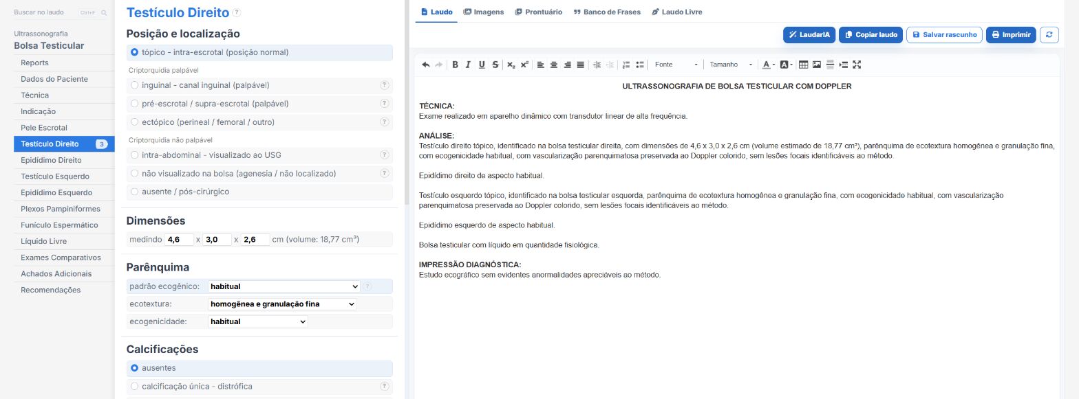
Keeping this standardization across all studies, especially on call shifts with patients referred from the emergency department and urology clinics, is the great practical challenge. Laudário was designed for exactly this day-to-day reality.
- Specific templates for Scrotum and Scrotum with Doppler, already structured in scanning order, with testes, epididymides, tunicae, spermatic cord, varicocele assessment and vascular evaluation.
- Ready-made fields for bilateral testicular dimensions and volume, with automatic calculation by the most widely used formulas and comparison between sides, avoiding manual math and transcription errors.
- Standardized, customizable phrases for the most frequent scenarios (normal study, left varicocele, suspected torsion, epididymo-orchitis, hydrocele, microlithiasis, testicular mass), keeping terminology consistent across reports for the same patient.
- The ability to describe associated findings (epididymal cyst, microlithiasis, small hydrocele) without leaving the same template, keeping typing flowing.
- Impression integrated with the description: the answer to the clinical question is built in parallel with the findings, avoiding inconsistencies between the body of the report and the closing.
- A complete library covering scrotum, prostate, kidneys and urinary tract, abdomen, thyroid, breast, pelvis, obstetrics and Doppler in general, all in a single online system.
- Professional presentation of the final report, with the clinic letterhead, electronic signature and a digital version with a validation QR Code, especially useful in emergency reports that need to circulate quickly among physicians and departments.
In practice, the time spent describing each structure, remembering every parameter and structuring the impression is reduced to a few clicks. Instead of starting from a blank page with every patient, you adjust a template that has already been tested and focus on what really matters: careful reading of the images and clinical interpretation of the findings.
If your practice frequently sees cases of acute scrotal pain, varicocele and male infertility workup, it is worth trying Laudário in practice. Try it free for 15 days, no credit card required, and within a few studies you will notice the difference that a system built for the ultrasound routine makes in report production.
The ultrasound reporting platform built by sonologists. We write about reports, classifications and standardization from the real routine of the practice. Who writes and how.
Educational content for healthcare professionals. It does not replace clinical judgment or the guidelines adopted by your service. Editorial policy.