Bladder Ultrasound: Volume, Postvoid Residual and What to Describe in the Wall
How to calculate bladder volume and postvoid residual, when the residual is significant, what to measure in the wall, trabeculation and diverticula, and the findings that call for cystoscopy.
 LaudárioUltrasound reports in practice
The bladder is included in almost every total abdomen, urinary tract and prostate ultrasound, and it is almost always described with the same sentence. But postvoid residual is one of the measurements urologists request most, and a poorly described wall thickening can be either a trabeculated bladder or a tumor. A useful report gives numbers and distinguishes diffuse from focal.
Bladder volume
Volume is calculated with the ellipsoid formula, from the three bladder measurements (longitudinal, transverse and anteroposterior), multiplied by a coefficient of about 0.52. The result is an estimate, with greater error in poorly filled or irregularly shaped bladders. The report should record the measurements and the calculated volume.
To examine the wall and the contents, the bladder needs to be well filled. An empty or poorly filled bladder is a technical limitation and should be mentioned.
Postvoid residual
The residual is measured right after voiding, ideally within a few minutes, with the same formula. There is no single cutoff accepted by all guidelines, but in practice:
- A small residual, below about 50 mL, is generally considered adequate in adults.
- Residuals above 100 mL are usually considered significant and deserve emphasis.
- Between 50 and 100 mL, the finding calls for clinical correlation and, if necessary, repeat measurement.
The report should state the prevoid volume and the residual, and whether there was a prolonged interval between voiding and the measurement, which overestimates the residual.
Bladder wall
- With the bladder full, the normal wall is thin, generally up to about 3 mm. Measurement in an empty bladder overestimates the thickness.
- Diffuse thickening with trabeculation and diverticula suggests a bladder under strain from bladder outlet obstruction (prostatic hyperplasia, urethral stricture) or a neurogenic bladder.
- Focal thickening or a vegetating lesion, especially with flow on Doppler, cannot be attributed to strain. The recommendation is urologic evaluation with cystoscopy.
Other findings
- Diverticula: number, location, neck and content; a diverticulum can hide a stone or a tumor.
- Stones: hyperechoic image with shadowing, which moves with a change in position.
- Clots and debris: echogenic material without shadowing and without flow on Doppler.
- Ureterocele: cystic dilatation of the intravesical portion of the ureter.
- Ureteral jets on color Doppler, useful when ureteral obstruction is suspected.
Common mistakes
- Residual without the prevoid volume, or measured long after voiding.
- Measuring wall thickness with an empty bladder.
- Attributing focal thickening to a "trabeculated bladder".
- Not using Doppler on a vegetating lesion.
- Omitting that the bladder was poorly filled.
See also our article on prostate ultrasound.
How Laudário helps
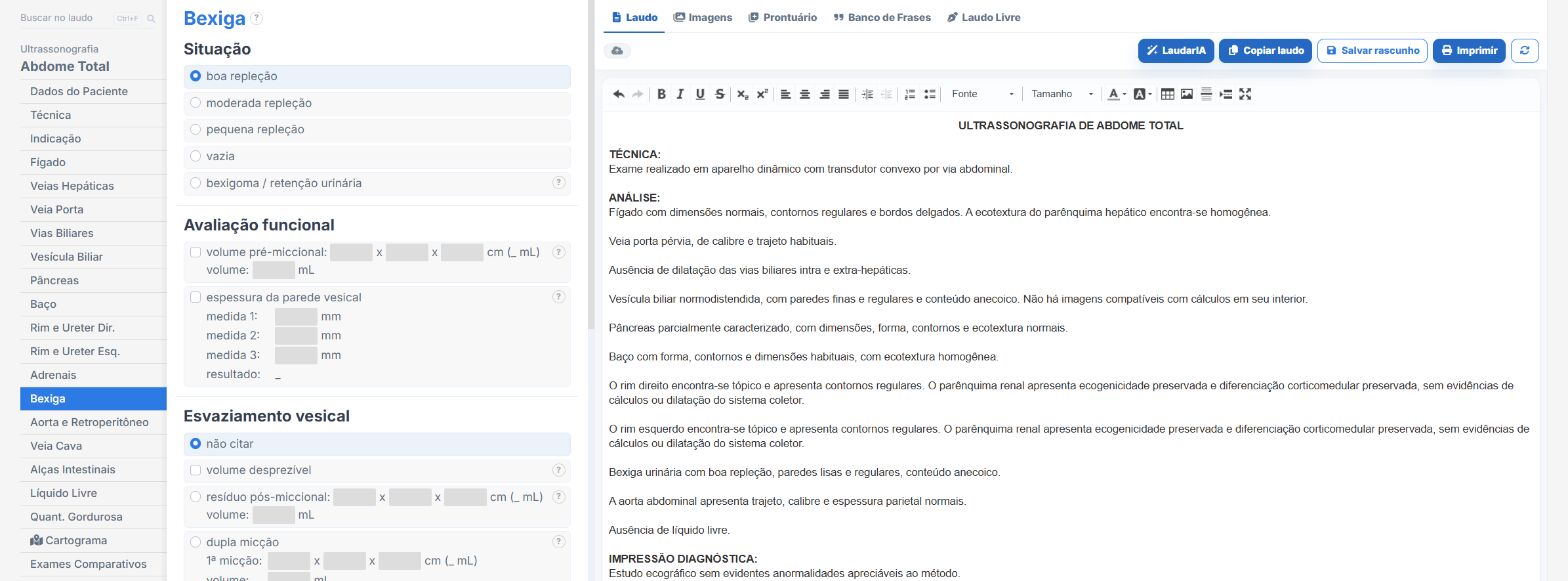
In the urinary tract and prostate templates of Laudário:
- Bladder volume and postvoid residual calculated from the measurements.
- Wall, diverticula and lesions in dedicated fields.
- Recommendations in their own tab.
Try it for 15 days free, no credit card.
The ultrasound reporting platform built by sonologists. We write about reports, classifications and standardization from the real routine of the practice. Who writes and how.
Educational content for healthcare professionals. It does not replace clinical judgment or the guidelines adopted by your service. Editorial policy.