Renal Cyst on Ultrasound: The Simple Cyst, the Bosniak Classification and When to Order CT
Which criteria define a simple renal cyst on ultrasound, why the Bosniak classification was built for CT and MRI, and how to report a cyst that is not simple without alarming and without missing anything.
 LaudárioUltrasound reports in practice
The renal cyst is one of the most common findings on abdominal ultrasound, and the great majority are simple and clinically unimportant. The problem lies with those that are not. A report that calls a lesion with a thick septum a "renal cyst", or that applies Bosniak to what ultrasound cannot classify, can either trigger an unnecessary workup or let a cystic carcinoma slip by.
The simple cyst
On ultrasound, the simple renal cyst meets all of these criteria:
- Anechoic content.
- Thin, smooth wall, barely perceptible.
- Round or oval shape, well defined.
- Posterior acoustic enhancement.
- No septa, calcifications, solid component or flow on Doppler.
When all criteria are met, the lesion is benign and needs no imaging follow-up. The report describes the side, the third of the kidney, the location (cortical, parapelvic, exophytic) and the largest measurement.
Bosniak: a CT and MRI classification
The Bosniak classification, revised in 2019, was built for contrast-enhanced CT and MRI, because it depends on enhancement of the wall, septa and solid components. Conventional ultrasound does not measure enhancement. Therefore, applying categories II, IIF, III and IV from B-mode is an extrapolation of the classification.
Contrast-enhanced ultrasound (CEUS) is able to assess enhancement and has been used to characterize complex cysts, but it is still not available in most services. On conventional ultrasound, the safe approach is to describe the findings and recommend the appropriate modality.
The cyst that is not simple
Any of these findings takes the cyst out of the simple category:
- Septa: thin or thick, single or multiple.
- Calcifications in the wall or septa.
- Content with echoes (hemorrhagic or proteinaceous), which may be benign but is not anechoic.
- Wall thickening or nodularity.
- Solid component, especially with flow on Doppler.
In these cases, the report describes what was seen and recommends contrast-enhanced CT or MRI for characterization. A vascularized solid component justifies highlighting the recommendation, because it is the finding most associated with malignancy.
Other situations
- A parapelvic cyst can be mistaken for hydronephrosis. The cyst does not communicate with the calyces; when in doubt, excretory-phase CT resolves it.
- Multiple bilateral cysts in a young patient raise the hypothesis of polycystic kidney disease; the report should count the cysts in each kidney and measure the kidneys.
- A cyst in the kidney of a patient on dialysis is part of acquired cystic disease, which carries a higher risk of neoplasia and deserves careful description.
Common errors
- Classifying by Bosniak from conventional ultrasound.
- Calling a lesion with a septum, calcification or echoes a simple cyst.
- Omitting Doppler in a cyst with a solid component.
- Recommending ultrasound follow-up for a cyst that needs CT.
- Failing to measure, which prevents comparison.
See also our text on the kidney and urinary tract report.
How Laudário helps
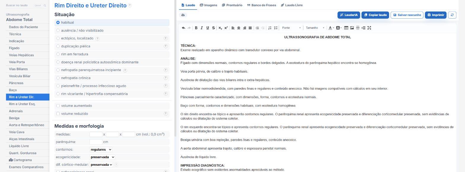
In the abdomen and urinary tract templates of Laudário:
- Renal cysts with side, location, measurements and characteristics in their own fields.
- The conclusion is assembled from the checked findings, with no retyping.
- Recommendations in their own tab, such as CT or MRI for characterization.
Try it for 15 days free, no credit card.
The ultrasound reporting platform built by sonologists. We write about reports, classifications and standardization from the real routine of the practice. Who writes and how.
Educational content for healthcare professionals. It does not replace clinical judgment or the guidelines adopted by your service. Editorial policy.