Hepatic Doppler and portal hypertension: how to structure the report, from portal flow to TIPS
Portal vein caliber is the weakest sign of the exam and may even decrease in advanced disease. See what really informs: flow direction, hepatic vein waveform, arterialization, and collaterals (the most specific finding of all), plus TIPS and transplant.
 LaudárioUltrasound reports in practice
This article is part of the guide Abdominal Ultrasound: A Guide to the Findings That Actually Change Management.
Portal hypertension is a hemodynamic diagnosis, and Doppler ultrasound is the only routine method capable of watching it happen. It is not a measurement: it is a set of signs that, read together, tell the story of a liver that has stopped accepting the blood the intestine sends it. The problem is that most hepatic Doppler reports list the signs in isolation, without assembling the story: "patent portal vein, caliber 1.4 cm" says nothing about portal pressure.
This text walks through the exam in the order in which it answers the clinical question: the portal vein and flow direction, the hepatic veins and their waveform, the hepatic artery and what it does when the portal vein fails, the collaterals (the most specific finding of all), and the two post-intervention scenarios that most often reach the clinic: TIPS and transplant.
The portal vein: caliber is the weakest sign, direction is the strongest
Start with what matters least, to get it out of the way. The portal vein caliber (usual reference up to about 1.2 to 1.3 cm at the hilum, in quiet inspiration) is the most cited parameter and the least useful: there is enormous overlap between normal and hypertensive patients, and, the counterintuitive point, it may even decrease in advanced portal hypertension, when collaterals divert the flow and the portal vein loses volume. A portal vein of normal caliber does not rule out portal hypertension; a dilated portal vein, in isolation, does not confirm it.
What really informs is the hemodynamics:
- Mean velocity: approximate reference of 16 to 40 cm/s. Slow flow is the first sign to appear.
- Respiratory phasicity: the normal portal vein oscillates with respiration. Loss of this phasicity, with monotonous flow, is an early and frequently ignored sign.
- Direction: and here is the strongest sign of the exam.
Direction has three states, in order of severity:
- Hepatopetal: toward the liver. The normal state.
- Bidirectional: alternating throughout the respiratory cycle. Severe portal hypertension.
- Hepatofugal: reversed, away from the liver. Advanced portal hypertension, with blood being drained through collaterals instead of crossing the parenchyma.
There is also the congestion index (cross-sectional area ÷ mean velocity, approximate reference ≤ 0.07), which combines both axes into a single number and rises early. It is little used in routine practice and is worth using.
Portal vein thrombosis: the distinction that changes everything
Faced with echogenic intraluminal content, there is one question the report must answer: does the thrombus have internal vascularity on Doppler?
- No internal flow: bland thrombus.
- With internal flow, typically pulsatile arterial flow within the thrombus: tumor thrombus until proven otherwise, and in the setting of cirrhosis this means hepatocellular carcinoma invading the portal vein.
This is an oncologic staging distinction made with color Doppler, and it transforms the report. Also record the extent (branch, trunk, or extending into the superior mesenteric vein): thrombosis extending into the SMV carries a risk of intestinal ischemia and is an immediately serious finding.
In chronic thrombosis, the portal vein is replaced by a tangle of tortuous low-velocity channels: cavernous transformation. The cavernoma is the fossil of an old thrombosis.
Hepatic veins: the waveform tells what lies downstream
The normal hepatic vein has a triphasic waveform, and each component has a name and meaning: S (antegrade systolic), D (antegrade diastolic), and A (retrograde presystolic, from atrial contraction). This waveform is a window into the right heart: it reflects the atrial pressure transmitted retrogradely.
Interpreting the changes is directional and has two opposite vectors:
Loss of phasicity (flattening): the waveform goes from triphasic to biphasic (loses the A wave) and then to monophasic, nearly continuous. This reflects loss of hepatic compliance: a stiff, cirrhotic liver no longer transmits the oscillations. It is the same phenomenon that liver elastography measures directly, which is why the two exams corroborate each other.
Increased pulsatility: the opposite: oscillations are amplified and the A wave becomes accentuated. This is not liver disease; it is passive congestion, the cardiac liver of right heart failure. And there is a recognizable progression:
- Flattened S wave, inverted S/D ratio: elevated right atrial pressure or mild to moderate tricuspid regurgitation.
- Reversed S wave (W pattern): hemodynamically significant tricuspid regurgitation.
- "To and fro" pattern, frankly bidirectional: severe right-sided congestive heart failure.
This is one of the most undervalued findings of abdominal Doppler: a pulsatile hepatic waveform on a routine exam is a cardiac diagnosis, and the report may be the first document to suggest evaluation of tricuspid function.
Absence of flow in the hepatic veins raises Budd-Chiari syndrome, and it is worth grading certainty by extent: one vein without flow is "suggestive"; two or more is "strongly suggestive." Supporting findings: caudate lobe hypertrophy (which has its own drainage to the cava and is therefore spared), intrahepatic collaterals, and a dilated caudate vein.
The hepatic artery: the liver compensating
The normal hepatic artery has a low-resistance waveform, with sustained positive diastolic flow, and a resistive index between 0.55 and 0.80. Usual peak systolic velocity is between 30 and 60 cm/s.
The concept that organizes everything here is the hepatic artery buffer response: when portal inflow falls, the artery compensates by increasing flow. A hyperemic hepatic artery, with elevated PSV (above ~100 cm/s), in a patient with slow portal flow is not an isolated abnormality: it is the liver trying to maintain perfusion. This is arterialization, and it tells the same story as the portal vein.
Two patterns deserve a name:
- Tardus-parvus: slow, rounded systolic upstroke of low amplitude, with a prolonged acceleration time (above ~80 ms). It means hemodynamically significant upstream stenosis, even if the stenosis is not seen directly. It is a powerful indirect sign.
- High resistance (RI above 0.80): nonspecific but relevant; it may reflect diffuse liver disease, biliary obstruction, or systemic hemodynamic change.
Direct signs of arterial stenosis: focal elevation of PSV (above ~200 cm/s), post-stenotic turbulence with aliasing, and a velocity gradient between segments above 3:1.
Collaterals: the most specific finding of the exam
If there is one block that separates a competent hepatic Doppler report from a generic one, this is it. A portosystemic collateral is the finding with the highest specificity for clinically significant portal hypertension, more than caliber, more than velocity, even more than slow flow. Blood that cannot cross the liver has to go somewhere, and it takes predictable paths:
- Recanalized paraumbilical vein: in the falciform ligament, in continuity with the left portal branch, with hepatofugal flow draining to the abdominal wall (Cruveilhier-Baumgarten syndrome, whose clinical expression is caput medusae). This is a highly specific finding, practically nonexistent without portal hypertension.
- Dilated left gastric (coronary) vein: above ~5 mm, in the lesser omentum, with reversed flow toward the azygos system. It is an independent marker of risk for esophagogastric varices, and therefore the finding that most directly indicates endoscopy.
- Esophagogastric varices: tubular, tortuous structures near the gastroesophageal junction and the gastric fundus.
- Spontaneous splenorenal shunt: serpiginous plexus at the splenic hilum draining into the left renal vein. A high-output decompression route, associated with advanced disease and encephalopathy.
- Perigastric and perisplenic collaterals (short gastric) and retroperitoneal varices (communicating with the azygos and hemiazygos).
A point of wording worth adopting: reserve the qualifier "clinically significant" for when a collateral is identified. Isolated slow flow suggests portal hypertension; slow flow with a collateral characterizes it as clinically significant. This is an honest gradation, and it speaks directly to the 17 kPa cutoff of elastography, which points to the same outcome by a different route.
The tributaries
They complete the map and often confirm the diagnosis when the portal vein is ambiguous:
- Splenic vein: caliber above ~0.9 cm suggests portal hypertension; so does flow reversal.
- Superior mesenteric vein: above ~1.2 cm. SMV thrombosis is a serious finding because of the risk of intestinal ischemia.
- Splenic artery: elevated RI suggests splenic congestion.
- Splenic artery aneurysm: worth looking for; the yin-yang sign on Doppler raises pseudoaneurysm, especially in the setting of pancreatitis.
And do not forget the spleen: splenomegaly is part of the picture and often falls outside the hepatic Doppler protocol, left "for the abdomen." Measure it.
TIPS: where hepatofugal becomes good news
The transjugular intrahepatic portosystemic shunt is a prosthesis that needs periodic surveillance, and Doppler is the method of choice. The references:
- PSV within the stent: 90 to 190 cm/s is the range of good function.
- Below 90 cm/s: possible onset of dysfunction; below 50 cm/s, severe dysfunction or occlusion.
- Above 190 cm/s: possible focal stenosis; above 250 cm/s, hemodynamically significant stenosis.
- A change of more than 50 cm/s from the previous exam is a warning sign, regardless of the absolute value. As in elastography, the delta matters more than the number, which is why the previous exam must be at hand.
And here is the most important conceptual reversal in this text: hepatofugal flow in the portal vein, in a patient with a TIPS, is an expected and good finding. It means the shunt is working and draining the portal system, exactly as designed. The same finding that, in the native liver, indicates advanced portal hypertension, here indicates therapeutic success. Reading a TIPS with the yardstick of the native liver produces alarming and wrong reports.
Liver transplant: the resistive index has its moment
In the graft, Doppler is the surveillance exam that detects vascular complications, and the most feared, hepatic artery thrombosis, is an emergency that can cost the graft.
Useful references:
- Intrahepatic RI: 0.55 to 0.80.
- RI below 0.50 with tardus-parvus: suspected critical proximal stenosis of the graft artery.
- RI above 0.85: nonspecific; possible causes include acute rejection, tubular necrosis, biliary obstruction, and veno-occlusive disease.
- PSV at the anastomosis above ~200 to 300 cm/s, or a gradient greater than 3:1 relative to the pre-anastomotic segment: stenosis.
One detail that avoids a false alarm: a slightly elevated RI (0.80 to 0.85) may be normal in the first 72 hours after surgery. This is the only point in the exam where time since surgery enters the interpretation, and a report that ignores it triggers unnecessary workup in a patient who is only on the second day.
Pitfalls of the hepatic Doppler report
- Concluding portal hypertension from portal vein caliber: it is the weakest sign, and it can decrease in advanced disease.
- Not recording the direction of portal flow: it is the strongest sign of the exam.
- Not checking vascularity within the thrombus: it separates bland thrombus from tumor invasion.
- Reading a pulsatile hepatic waveform as liver disease: it is cardiac congestion, and the referral is different.
- Not looking for collaterals: the most specific finding of the exam, and the one that indicates endoscopy.
- Applying the native liver yardstick to a TIPS: hepatofugal there is success, not catastrophe.
- Evaluating a TIPS without the previous exam: the 50 cm/s delta is the criterion, and it requires the series.
- Being alarmed by an RI of 0.82 on the second day after transplant: it may be physiological in the first 72 h.
- Forgetting the spleen: splenomegaly is part of the picture.
- Delivering numbers without the synthesis: the report must say whether the whole configures portal hypertension, not just list velocities.
How Laudário helps structure the hepatic Doppler
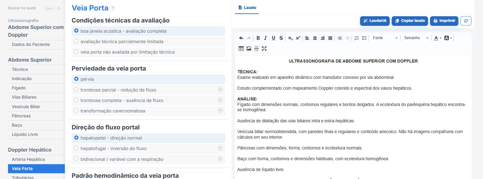
Laudário has a module dedicated to hepatic Doppler, with tabs in the order of hemodynamic reasoning and the two special situations as optional blocks. In practice:
- Reference value displayed next to each measurement, with automatic classification (normal, reduced, increased): portal caliber and velocity, congestion index calculated automatically from the two, splenic and SMV calibers, and arterial RI derived from the velocities.
- Automatic synthesis of portal hypertension: the system builds the conclusion by listing the signs actually present (slow flow, reversal, collaterals) and reserves "clinically significant" for when a collateral is identified.
- The seven portosystemic collaterals as their own fields, with complete phrases, including Cruveilhier-Baumgarten and splenorenal shunt, and automatic aggregation according to the number found.
- The ten hepatic vein waveform patterns, with the semiology of each written out: biphasic, monophasic, flattened S, W pattern, to and fro.
- TIPS block with automatic classification by maximum PSV and calculation of the delta against the previous exam, with an alert above 50 cm/s.
- Transplant block with calculated mean RI, five classification ranges, and the caveat for the first 72 hours.
- Auto-suggested recommendations that close the clinical loop: endoscopy when there is portal hypertension with decompression, echocardiography with targeted tricuspid evaluation when the pattern is congestive, thrombophilia workup in the face of thrombosis.
- Support from LaudarIA for cases in which the findings contradict each other.
If you perform hepatic Doppler and build the report manually, it is worth trying: 15 days of free trial, no credit card required, with the hepatic modules (Doppler, elastography, fat quantification, and multiparametric assessment) all unlocked.
The ultrasound reporting platform built by sonologists. We write about reports, classifications and standardization from the real routine of the practice. Who writes and how.
Educational content for healthcare professionals. It does not replace clinical judgment or the guidelines adopted by your service. Editorial policy.