Focal liver lesions on ultrasound: cyst, hemangioma, FNH, metastasis and what to write in the report
How to recognize the simple cyst and the typical hemangioma, when a nodule calls for CT or MRI, what changes in the oncology and cirrhosis patient, and what must not be missing from the description.
 LaudárioUltrasound reports in practice
This article is part of the guide Abdominal Ultrasound: A Guide to the Findings That Actually Change Management.
A liver nodule found by chance is one of the findings that generates the most anxiety for patients and rework in the office. The good news is that many of them have a typical ultrasound appearance and can be described with confidence. What changes management is the context: the same hyperechoic nodule means different things in a healthy adult, in an oncology patient and in a cirrhotic patient.
Simple cyst
Anechoic, with thin, imperceptible walls, regular contours and posterior acoustic enhancement. With these criteria, it is benign and needs no follow-up. Thick septa, a solid component, a mural nodule or flow on Doppler take the cyst out of the simple category and call for evaluation with computed tomography (CT) or magnetic resonance imaging (MRI).
Hemangioma
It is the most common benign liver tumor. The typical appearance is a hyperechoic, homogeneous, well-defined nodule, usually small, sometimes with mild posterior enhancement and no significant central flow on Doppler.
- In a patient without chronic liver disease and without known malignancy, a small typical hemangioma is usually accepted as a diagnosis on ultrasound.
- Large, heterogeneous or atypical-appearing hemangiomas, and any hyperechoic nodule in an oncology patient, should be confirmed by contrast-enhanced CT or MRI.
Focal nodular hyperplasia and adenoma
- Focal nodular hyperplasia (FNH): often isoechoic to the parenchyma, noticed more by its effect on the vessels than by its echogenicity. It may have a central scar and radiating flow on Doppler, in a "spoke-wheel" pattern.
- Hepatocellular adenoma: more common in women using hormonal contraceptives; it may be heterogeneous due to bleeding. It carries a risk of hemorrhage and, in some subtypes, of malignant transformation.
Neither is reliably confirmed on ultrasound. The report describes the nodule and recommends characterization by MRI.
Focal steatosis and focal sparing
In a fatty liver, spared areas appear as hypoechoic regions; in a normal liver, focal fatty infiltration appears hyperechoic. Typical locations are next to the gallbladder, the porta hepatis and the falciform ligament. What helps: a geographic shape, absence of mass effect and vessels crossing the area without displacement.
Metastases
They are usually multiple, of variable echogenicity, and a peripheral hypoechoic halo (the "target" pattern) is a warning sign. In an oncology patient, any new nodule deserves comparison with prior exams and, in general, characterization by CT or MRI.
Cirrhotic liver: a different line of reasoning
In a patient with cirrhosis or chronic hepatitis B under surveillance, a nodule is not presumed benign. By ultrasound LI-RADS, an observation of 10 mm or more, or new portal vein thrombus, classifies the exam as positive (US-3) and indicates multiphase CT or MRI. Nodules smaller than 10 mm go into closer ultrasound follow-up.
What to describe
- Number of lesions and hepatic segment of each one (Couinaud classification).
- Dimensions, echogenicity, contours and presence of a halo.
- Behavior on Doppler.
- Relationship to vessels and to the capsule.
- Comparison with prior exams: stability is valuable information.
- Context that changes management: chronic liver disease, known malignancy.
Common errors
- Calling a hyperechoic nodule a hemangioma in an oncology patient without recommending characterization.
- Omitting the hepatic segment, which makes follow-up harder.
- Reporting an area of focal sparing in steatosis as a nodule.
- Applying to the cirrhotic liver the same reasoning used for the normal liver.
See also the texts on hepatic steatosis and liver elastography.
How Laudário helps
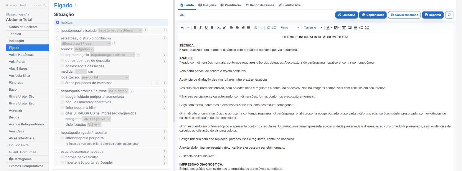
In the upper abdomen and total abdomen templates of Laudário:
- Liver lesions with segment, measurements, echogenicity and Doppler in their own fields.
- Numbered nodule adder, one per lesion, for follow-up.
- Recommendations in their own tab, separate from the findings.
Try it for 15 days free, no credit card required.
The ultrasound reporting platform built by sonologists. We write about reports, classifications and standardization from the real routine of the practice. Who writes and how.
Educational content for healthcare professionals. It does not replace clinical judgment or the guidelines adopted by your service. Editorial policy.