Soft Tissue Ultrasound: How to Structure the Report for Subcutaneous Nodules Clearly
How to structure the soft tissue ultrasound report, describe subcutaneous nodules, lipomas, cysts and collections clearly, and standardize the diagnostic impression.
 LaudárioUltrasound reports in practice
Soft tissue ultrasound is a versatile and often underestimated exam: it shows up every day in office practice, the emergency department and the imaging clinic, yet it rarely gets a report template as structured as exams like the thyroid or breast. The result is reports that are too short, such as “hypoechoic nodular image in the cervical region, no signs of inflammation”, which do little to help the referring physician decide between observing, aspirating or referring. In this article, we lay out a practical structure for the soft tissue ultrasound report, focusing on subcutaneous nodules, collections and body wall lesions.
Why are soft tissue reports often poor?
The exam is technically simple, but the range of findings is enormous: lipomas, epidermal cysts, abscesses, hematomas, reactive lymph nodes, abdominal wall hernias, schwannomas, soft tissue tumors and vascular lesions. Because there is no single, widely accepted classification for this territory (such as BI-RADS or TI-RADS), many professionals end up describing findings generically, without objective criteria.
The main problems in this scenario are:
- Lack of precise location: “right arm region” is not enough. The referring physician needs to know which third, which surface and which plane (cutaneous, subcutaneous, intramuscular).
- Incomplete description of the lesion: dimensions in a single plane, with no echogenicity, margins or vascularity, leave the report open to interpretation.
- No diagnostic hypothesis: describing without suggesting a hypothesis (lipoma, cyst, abscess, lymph node) reduces the clinical value of the report.
- Confusion between solid, cystic and mixed lesions: a wrong classification at this point can lead to mistaken management, such as aspirating a typical lipoma or merely following a collection that needs to be drained.
Recommended structure for the soft tissue ultrasound report
The logic of the report should follow clinical reasoning: locate the lesion precisely, describe its morphologic attributes, assess vascularity and correlate with the most likely diagnostic hypothesis. We suggest the following blocks:
1. Identification and technique
Include the exam type (soft tissue ultrasound), the region evaluated according to the clinical request, the equipment (when relevant) and any technical limitation: patient body habitus, presence of a dressing, pain on compression, areas that are hard to access. When the exam is directed at a specific complaint (“palpable nodule in the right lateral cervical region”), reproduce the complaint in the report to give context to the reading.
2. Anatomic location
This is one of the most neglected points. Each lesion should be located with:
- Body region and laterality: right lateral cervical, left inguinal region, right back, middle third of the left arm.
- Depth plane: cutaneous, subcutaneous, fascial, intramuscular or periosteal.
- Relationship with neighboring structures: contact with vessels, nerves, fasciae, bones or joint planes.
- Distance from the skin: useful for lesions that will be aspirated or resected (especially for the surgeon).
3. Morphologic description of the lesion
For each relevant lesion, describe systematically:
- Dimensions in three planes (longitudinal, transverse and anteroposterior) whenever possible, with the unit stated clearly (mm or cm).
- Composition: solid, cystic (anechoic), mixed cystic and solid, or predominantly fatty.
- Echogenicity: hyperechoic, isoechoic, hypoechoic, anechoic or heterogeneous, always relative to the adjacent subcutaneous tissue or muscle.
- Margins: well defined, lobulated, ill defined, infiltrative.
- Capsule: present, absent or partial.
- Posterior acoustic enhancement, acoustic shadowing or characteristic artifacts.
- Compressibility and mobility on transducer pressure, especially useful in lipomas and cysts.
4. Doppler assessment
Color Doppler (and, when available, power Doppler) is decisive for differentiating lesions. Describe whether vascularity is absent, peripheral, central, diffusely increased, or shows a pattern suggestive of anomalous arterial or venous flow. In collections and abscesses, comment on perilesional hyperemia. In lymph nodes, assess the hilar versus peripheral or chaotic vascular pattern.
5. Associated findings
Do not treat the lesion as isolated. Comment on edema of the adjacent subcutaneous tissue, skin thickening, intact or blurred fascial planes, enlarged satellite lymph nodes and other changes in the region examined. In inflammatory conditions, this context is as valuable as the description of the collection itself.
6. Diagnostic impression and suggested management
The conclusion should be objective, with a main diagnostic hypothesis and, when appropriate, differential diagnoses. When the finding is classic (a typical lipoma, an uncomplicated epidermal cyst, a reactive lymph node), say so clearly instead of handing a vague description back to the clinician. When there is doubt or a finding suspicious for a soft tissue neoplasm, recommend clinical correlation and, if necessary, complementary magnetic resonance imaging or specialist evaluation.
Common findings and how to describe them
Lipoma
Solid lesion, usually subcutaneous, oval, with its long axis parallel to the skin, isoechoic or slightly hyperechoic relative to the subcutaneous tissue, with internal echogenic striations, no significant vascularity on Doppler and compressible with the transducer. Mentioning the depth and the planes involved is especially useful for the surgeon.
Epidermal cyst
Well-circumscribed nodular image, predominantly subcutaneous, hypoechoic or heterogeneous, with posterior acoustic enhancement, often with an extension to the skin (the “punctum” sign) and no internal vascularity on Doppler. In ruptured or inflamed cases, describe the infiltration of the adjacent subcutaneous tissue and the perilesional hyperemia.
Abscess and collection
Hypoanechoic, heterogeneous image with mobile echogenic debris, no central flow on Doppler, but with perilesional hyperemia. Reporting precise dimensions, depth and relationship with neighboring planes is essential for the team that will decide on drainage.
Reactive versus suspicious lymph node
A reactive lymph node tends to be oval, with a preserved echogenic hilum, homogeneous cortex and central hilar vascularity. A suspicious lymph node, in contrast, tends to be rounded (short-to-long axis ratio > 0.5), with loss or displacement of the hilum, focal cortical thickening and chaotic peripheral vascularity. Standardizing these phrases avoids ambiguous interpretations.
Abdominal wall hernia
In wall ultrasound for hernia assessment, describe the hernial orifice (location and largest diameter), the herniated content (fat, bowel loop), the behavior at rest and during the Valsalva maneuver, and whether or not it is reducible. The Valsalva maneuver must be stated explicitly in the report.
Common mistakes in the soft tissue report
- Generalizing the location: “cervical region” with no laterality or level, or “lower limb” with no third or surface.
- Not measuring in three planes: compromises follow-up, especially for lesions under surveillance.
- Ignoring Doppler: in collections, lymph nodes and tumors, vascularity completely changes the hypothesis.
- Not closing a diagnostic hypothesis: stopping at description hands the entire morphologic interpretation to the clinician.
- Not comparing with prior exams when available, especially for lesions in follow-up.
- Forgetting the Valsalva maneuver in ultrasound directed at an abdominal wall hernia.
How Laudário helps structure soft tissue reports
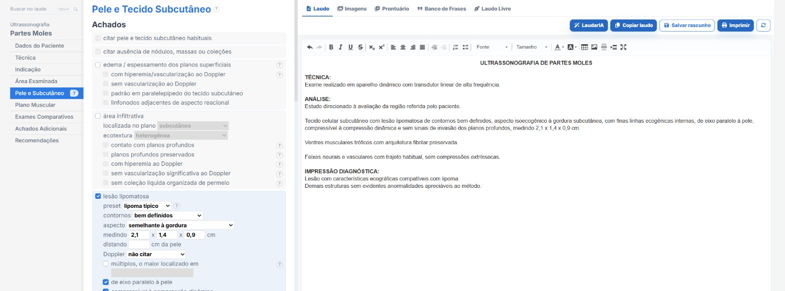
Laudário was designed precisely for this kind of scenario: an exam with many possible findings and little standardization between professionals. For soft tissue ultrasound and superficial exams in general, the platform offers:
- Ready-made, structured templates for soft tissues, abdominal wall, hernias, lymph nodes, joints and other superficial regions, organized in scanning order to speed up typing.
- Standardized, customizable phrases for describing lipomas, cysts, abscesses, and reactive and suspicious lymph nodes, keeping terminology consistent across reports and among the professionals of the same clinic.
- Automatic calculations and smart fields integrated into the workflow, avoiding manual math and reducing transcription errors in measurements and axis ratios.
- A complete library covering thyroid, breast, abdomen, pelvis, obstetrics, Doppler and soft tissue exams, all in a single online system.
The result is straightforward: less time typing, fewer forgotten findings and more consistent reports across exams of the same patient, which is essential in an area where following up an “indeterminate” lesion depends on comparing old descriptions with current ones. Instead of starting from scratch with each patient, you adjust a template that has already been tested, keep the clinic's professional standard and focus on what really demands your attention: reading the images.
If you still build your soft tissue reports manually or by reusing old text, it is worth getting to know Laudário. Try it free for 15 days and see in practice how a good structure saves time on every exam.
The ultrasound reporting platform built by sonologists. We write about reports, classifications and standardization from the real routine of the practice. Who writes and how.
Educational content for healthcare professionals. It does not replace clinical judgment or the guidelines adopted by your service. Editorial policy.