Abdominal wall hernia ultrasound: how to structure the report and avoid ambiguous findings
How to standardize the abdominal wall hernia ultrasound report: Valsalva technique, description of the defect, hernia contents and suggested management.
 LaudárioUltrasound reports in practice
Abdominal wall hernia ultrasound is a seemingly simple exam that hides several pitfalls in the report. The region is dynamic, the defect may appear only with maneuvers, the hernia contents change during the sweep, and terminology varies widely among practitioners. The result is a report that is hard to compare with the prior study and, often, of little use for the surgical decision. In this article, we lay out a practical structure for describing abdominal wall hernias with clarity and standardization.
Why does the hernia report demand more attention than it seems?
Unlike other soft tissue studies, evaluation of the abdominal wall only makes sense when it integrates anatomy, respiratory dynamics and maneuvers that increase intra-abdominal pressure. A real defect can go unnoticed at rest, and a vaguely described finding can lead the surgeon to indicate, or rule out, a repair without sufficient information.
The most common errors in this indication are:
- Not performing dynamic maneuvers: small hernias only manifest during Valsalva, coughing or standing, and the report needs to document the maneuver used.
- Confusing hernia with rectus diastasis: diastasis is widening of the linea alba, without an aponeurotic defect; treating the two as synonyms compromises management.
- Describing only the presence, without measuring the defect: the size of the neck is one of the main determinants of the surgical technique.
- Omitting the hernia contents: bowel loop, preperitoneal fat or omentum change the clinical meaning of the finding.
- Not commenting on reducibility: reducible, irreducible, incarcerated or strangulated hernias are situations of very different severity.
Recommended structure for the abdominal wall hernia ultrasound report
A good structure for this indication divides the wall into segments, organizes the description of each defect in a standardized way and closes with an objective conclusion. We suggest the following blocks:
1. Identification and technique
State the type of study (abdominal wall ultrasound for hernia assessment), the equipment used (when relevant), the transducer (usually a high-frequency linear, with a convex as an adjunct in patients with a thicker panniculus) and, above all, the maneuvers performed: quiet breathing, Valsalva, coughing and, when possible, standing. This block protects the practitioner and gives context to the referring physician.
2. Assessment by region
Instead of describing a single isolated finding, it is worth surveying the wall systematically. For each region, mention aponeurotic integrity, muscle thickness and the presence of defects:
- Epigastric region and linea alba: evaluation of the linea alba above the umbilical scar, with measurement of rectus diastasis at rest and during Valsalva.
- Umbilical and periumbilical region: integrity of the umbilical ring, contents of the scar and adjacent defects.
- Infraumbilical and suprapubic region: evaluation of the linea alba below the umbilicus and of the suprapubic region, including Spiegel hernias when suspected (lateral border of the rectus sheath).
- Inguinal and femoral regions: bilateral inguinal canal with assessment for indirect, direct and femoral hernia; describe the course of the spermatic cord and its relationship to the inferior epigastric vessels.
- Prior scars: whenever there is a surgical history, examine the scar area to rule out incisional hernia, even if this is not the main complaint.
3. Individual description of each defect
Each identified defect deserves its own paragraph, in the following sequence:
- Precise location: distance from the umbilical scar or another stable anatomical landmark, side (right/left) and plane.
- Hernia type: umbilical, epigastric, indirect inguinal, direct inguinal, femoral, Spiegel, lumbar, incisional or of another location.
- Dimensions of the defect (neck): measured in at least two axes, at rest and during Valsalva. This is decisive for the surgical technique.
- Hernia contents: preperitoneal fat, omentum, bowel loop (with or without identified peristalsis), fluid, or bladder in large inguinal hernias.
- Dynamic behavior: defect present only with Valsalva, when standing, or already at rest; spontaneous reducibility or with an external maneuver.
- Signs of complication: irreducible contents, wall edema, absence of peristalsis in the herniated loop, altered vascularity on Doppler.
When there are multiple defects (common in patients with prior surgeries), standardize a numbering (Defect 1, Defect 2) with a specific location, to make comparison on future studies easier.
4. Rectus diastasis
Assess the inter-rectus distance at three standardized levels: 3 cm above the umbilicus, at the level of the umbilicus and 2 cm below it, at rest and during Valsalva. Clearly distinguish diastasis (without an aponeurotic defect) from linea alba hernia (with a real defect). The most commonly used cutoffs consider significant widening to be above 22 mm in the supraumbilical region, but comparison with the prior study and the clinical context (postpartum, weight gain) usually weigh more than the isolated number.
5. Diagnostic impression and suggested management
The conclusion should be objective, hierarchical and correlated with the descriptive body. For each hernia, state the location, neck size, contents, reducibility and, when applicable, a suggestion for further workup (evaluation by a general surgeon, CT in selected cases, ultrasound follow-up). The surgical decision always belongs to the treating physician, but the report needs to deliver all the elements for that decision.
Common errors in the abdominal wall hernia report
Even with a defined structure, some recurring errors undermine the quality of these reports:
- Describing a “bulge” without defining whether there is an aponeurotic defect: a bulge may be a hernia, diastasis or simple muscle protrusion; the report needs to clarify.
- Not measuring the neck of the defect: the size of the hernial ring is the data point with the greatest impact on surgical management.
- Forgetting the contralateral side: in inguinal hernias, bilateral evaluation is part of the protocol, even when the patient reports a complaint on one side only.
- Not documenting maneuvers: a report of “no evidence of hernia” loses value if it does not say whether Valsalva and standing were performed.
- Poor comparisons: “stable compared with prior” without restating the measurements of the previous defect makes follow-up harder.
- Mixing terminologies: alternating between “umbilical hernia” and “umbilical scar hernia”, or using “incisional hernia” for any defect near a scar, creates noise.
How Laudário helps structure abdominal wall hernia reports
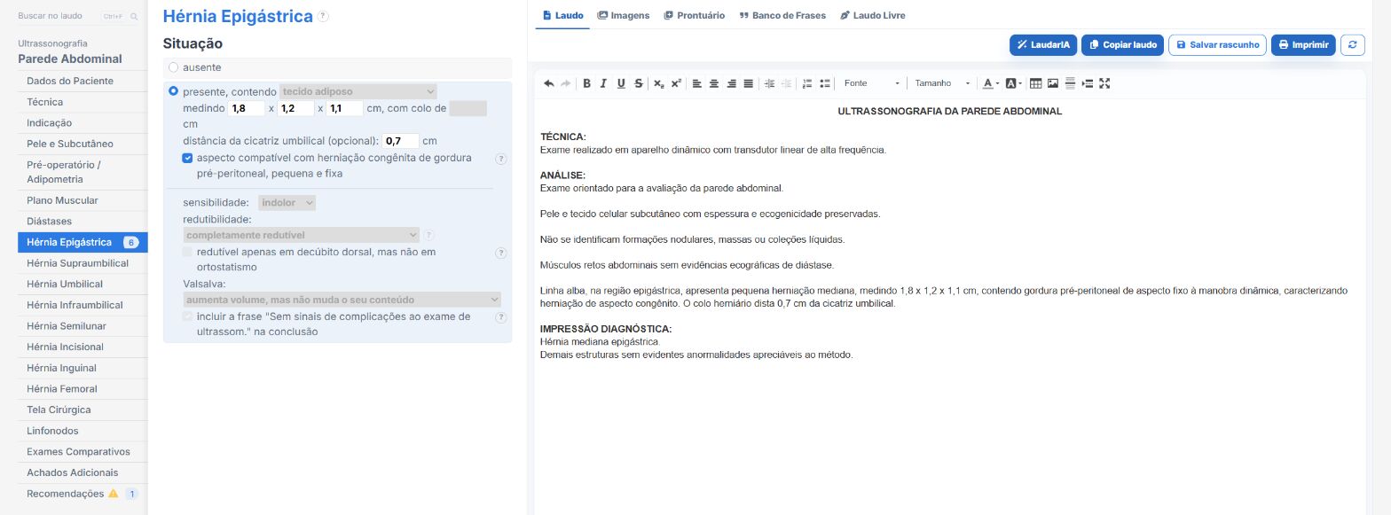
Laudário was designed for professionals who face this routine every day. For abdominal wall hernia ultrasound, and for the main soft tissue and abdominal protocols, the system offers:
- Ready-made, structured templates for abdominal wall, inguinal hernias, umbilical hernias, incisional hernias and rectus diastasis, organized in the order of the sweep to speed up typing.
- Standardized, customizable phrases for describing the defect, hernia contents, reducibility and dynamic behavior with Valsalva, keeping terminology consistent across reports.
- Dynamic fields for neck dimensions, distance from the umbilical scar and diastasis measurements at standardized points, avoiding omissions in the flow of the exam.
- A complete library covering abdomen, soft tissue, breast, thyroid, pelvis, obstetrics and Doppler studies, all in one online system.
The result is simple: less time typing, fewer forgotten findings and more consistent reports across studies of the same patient, which makes all the difference to the surgeon who will plan the repair. Instead of starting from a blank page with every patient, you adjust an already tested template, keep the practice's professional standard and focus on what really requires your attention: reading the images and integrating with the clinical picture.
If you still build your hernia reports manually or by copying old text, it is worth getting to know Laudário. Try it free for 15 days and see in practice how a well-designed structure saves time on every exam.
The ultrasound reporting platform built by sonologists. We write about reports, classifications and standardization from the real routine of the practice. Who writes and how.
Educational content for healthcare professionals. It does not replace clinical judgment or the guidelines adopted by your service. Editorial policy.