Abdominal aorta ultrasound: how to structure the report for aneurysm screening and assessment of dilatations
Abdominal aorta ultrasound is the method of choice for screening for abdominal aortic aneurysm (AAA) and following known dilatations. See how to structure the report with standardized suprarenal and infrarenal measurements, mural thrombus, common iliac arteries and the suggested management, and how Laudário speeds this up.
 LaudárioUltrasound reports in practice
Abdominal aorta ultrasound is a low-cost, radiation-free, highly accurate exam, considered the method of choice for screening for abdominal aortic aneurysm (AAA) and for following known dilatations. Although technically simple in most patients, the exam carries considerable clinical responsibility: AAA is a silent condition, often asymptomatic until rupture, and a well-structured report is what connects the sonographic finding to the vascular surgeon's therapeutic decision. Standardizing the description of diameters, mural thrombus, plaques and common iliac arteries is what separates a truly useful exam from a report that merely states “aorta of preserved caliber”.
Why standardize the abdominal aorta report
AAA screening is one of the few ultrasound indications with benefit proven at the population level in randomized trials, with a documented reduction in rupture mortality in men over 65, especially current or former smokers. For that benefit to translate into practice, the report must be objective: the vascular surgeon decides on follow-up, risk factor control or surgical indication from a single number (the maximum aortic diameter) and from how it is described.
Without standardization, the same exam can receive completely different reports: one describes only the infrarenal diameter, another measures on an oblique cut, another includes the thrombus in the measurement, and the next exam uses a different methodology, making comparison over time impossible. A standardized report describes the suprarenal and infrarenal segments, records diameters on a truly perpendicular cut, characterizes mural thrombus when present, evaluates the common iliac arteries and concludes by explicitly guiding management based on the largest diameter found.
Indications and data that should appear in the identification
The report should state the reason for the exam right at the start, because it completely changes what will be emphasized in the description and in the conclusion. The most common indications include screening in an asymptomatic patient with a risk factor (man over 65, smoker, family history of AAA), investigation of a pulsatile abdominal mass, abdominal or low back pain of indeterminate cause in a patient at risk, follow-up of a previously diagnosed aneurysm, and surveillance after endovascular or surgical repair.
When the exam is serial, it is worth recording the date of the previous study and explicitly stating whether the comparison shows stability, progression or a reduction in caliber. In patients who have undergone endovascular repair (EVAR), the indication should state this clearly, because the exam then focuses on looking for endoleak and on the behavior of the aneurysm sac over time, a completely different analysis from screening a treatment-naive patient.
Anatomy and technique: what cannot be missing
The abdominal aorta extends from the diaphragmatic hiatus to the iliac bifurcation, at approximately the level of the umbilicus, and can be divided into three segments with distinct practical implications for the report:
- Suprarenal: from the diaphragm to the origin of the renal arteries. Usually measuring 20 to 25 mm in adults. Aneurysms in this segment are less common but more complex surgically.
- Juxtarenal/pararenal: a short segment at the level of the renal arteries, important for surgical planning, since it determines whether there is an adequate proximal neck for endovascular repair.
- Infrarenal: the segment most often affected by AAA, below the renal arteries down to the bifurcation. More than 90% of abdominal aortic aneurysms occur here.
Proper technique begins with the patient fasting for 6 to 8 hours, supine, with a 2 to 5 MHz convex transducer. Measurements should be made on a truly perpendicular cut to the long axis of the vessel, in the anteroposterior (AP) diameter, including mural thrombus when present, from outer wall to outer wall (outer-to-outer). Measurements on an oblique cut overestimate the diameter and produce falsely positive reports for dilatation. In patients with an unfavorable body habitus or interposed bowel gas, gradual compression with the transducer and a change of position (left lateral decubitus) help displace bowel loops and improve the acoustic window.
Diagnostic criteria: when the aorta is aneurysmal
The diagnosis of abdominal aortic aneurysm is, by a convention widely accepted in the vascular literature, defined by a maximum anteroposterior diameter equal to or greater than 3.0 cm. Aortas between 2.5 and 2.9 cm are considered ectatic and should be described as such in the report, with a suggestion for follow-up within a defined timeframe according to the clinical context. Below 2.5 cm, the aorta is classified as normal caliber.
Once AAA is diagnosed, stratification by diameter range guides the follow-up interval and the surgical indication, according to the recommendations of the main vascular surgery societies:
- 3.0 to 3.9 cm: annual ultrasound surveillance.
- 4.0 to 4.9 cm: surveillance every 6 to 12 months, with specialist evaluation.
- 5.0 to 5.4 cm: surveillance every 3 to 6 months; formal surgical evaluation, especially in women.
- ≥ 5.5 cm in men or ≥ 5.0 cm in women: classic threshold for elective surgical indication.
- Growth > 0.5 cm in 6 months or > 1.0 cm in 1 year: surgical indication regardless of absolute diameter.
These cutoffs are widely used references in clinical practice and should appear, even if briefly, in the conclusion of the report, with the appropriate management suggestion. The sonologist does not replace the vascular surgeon, but a report that merely states “aorta with a diameter of 5.2 cm” without commenting on the clinical significance of the finding delivers less value than it could.
How to describe the aneurysm in the report
Once the dilatation is identified, the description must be complete enough for the vascular surgeon to decide between follow-up, complementary CT angiography or surgical indication without having to repeat the exam. The minimum elements are:
- Location: suprarenal, juxtarenal or infrarenal. For infrarenal AAA, state the approximate distance between the start of the dilatation and the origin of the renal arteries (proximal neck) and the distance between the end of the aneurysm and the aortic bifurcation (distal neck), information that is decisive for planning a possible EVAR.
- Shape: fusiform (more common) or saccular (less common, with a higher risk of rupture for the same diameter).
- Maximum anteroposterior diameter: in millimeters, on a perpendicular cut, from outer wall to outer wall, including mural thrombus when present.
- Transverse diameter: when relevant, to characterize the shape.
- Longitudinal extent: in millimeters, from the start to the end of the dilatation.
- Mural thrombus: presence, maximum thickness, location (anterior, posterior, lateral wall) and characteristics (homogeneous, heterogeneous, with areas of fissuring).
- Parietal calcifications: presence, distribution and relationship to possible future endovascular access sites.
- Signs of complication: focal wall irregularity, discontinuity of the inner layer suggesting dissection, signs of contained rupture (rare and typically identified on CT, but which can be suspected at ultrasound).
Accompanying findings: what must not be missed
Abdominal aorta ultrasound is rarely requested as an isolated exam outside the screening routine without the sonologist taking advantage of the window to evaluate neighboring structures. Even when the focus is strictly aortic, some accompanying findings must be actively sought, because they directly change management:
- Common iliac arteries: should be measured bilaterally, with AP diameter. An aneurysm is considered present when > 1.5 cm. Iliac aneurysms coexist with AAA in a significant proportion of cases and influence surgical planning.
- Atheromatous plaques of the aorta: describe thickness, extent and signs of instability (irregularity, ulceration).
- Aortic dissection: when identified, describe the presence of an intimal flap, false lumen, Doppler flow in both lumens and the relationship to the visceral branches. Ultrasound is less sensitive than CT for this diagnosis, but the finding mandates immediate complementary investigation.
- Inferior vena cava: describe caliber, inspiratory collapse and absence of masses or thrombi. Extrinsic compression by the dilated aorta can occur in large AAAs.
- Retroperitoneal lymphadenopathy: retroperitoneal fibrosis and periaortic lymphadenopathy can mimic inflammatory aneurysms and should be described.
Conclusion and clinical guidance
The conclusion of the abdominal aorta report should summarize, in a few lines, three essential pieces of information for the requesting physician: the largest diameter recorded in millimeters, the segment involved and the suggested management based on the follow-up ranges. When the exam is normal, an objective conclusion (“abdominal aorta of preserved caliber, with maximum AP diameter of XX mm in the infrarenal segment, without evidence of aneurysmal dilatation”) is more valuable than generic descriptions such as “aorta without abnormalities”.
In patients under surveillance, the conclusion must state the comparison with the previous exam: dimensional stability, reduction or progression. Simply repeating the diameter without contextualizing the comparison is a common failure that substantially reduces the clinical value of a serial report.
Findings that deserve emphasis in the conclusion
- Maximum AP diameter equal to or greater than 3.0 cm, with the corresponding range and a suggested surveillance interval.
- Significant growth compared with the previous exam (> 0.5 cm in 6 months or > 1.0 cm in 1 year), with an explicit suggestion for surgical evaluation.
- Saccular aneurysm, even below the elective treatment thresholds for fusiform aneurysms.
- Extensive or heterogeneous mural thrombus, or with signs of fissuring.
- Associated common iliac aneurysm, with diameter and laterality.
- Suspected dissection, contained rupture or inflammatory aneurysm, with an explicit indication for complementary CT angiography.
- Significant technical limitation due to bowel gas or body habitus, when the exam did not allow a reliable measurement: transparency here avoids false negatives.
Precautions to avoid ambiguous reports
- Always measure on a truly perpendicular cut to the long axis of the aorta; measurements on an oblique cut overestimate the diameter.
- Include the mural thrombus in the AP diameter measurement, from outer wall to outer wall, following the convention of the vascular societies.
- Record the diameter in millimeters, not in fractions of a centimeter with inconsistent decimal places.
- Distinguish aneurysmal dilatation (≥ 3.0 cm) from ectasia (2.5 to 2.9 cm); avoid using “mild dilatation” without the objective number.
- Evaluate and measure the common iliac arteries bilaterally, even when the aorta is normal: isolated iliac aneurysms exist and are clinically relevant.
- In follow-up exams, explicitly compare with the previous study; it is not enough to repeat the description.
- When the acoustic window is poor, record the technical limitation instead of forcing an unreliable measurement.
How Laudário helps structure the abdominal aorta report
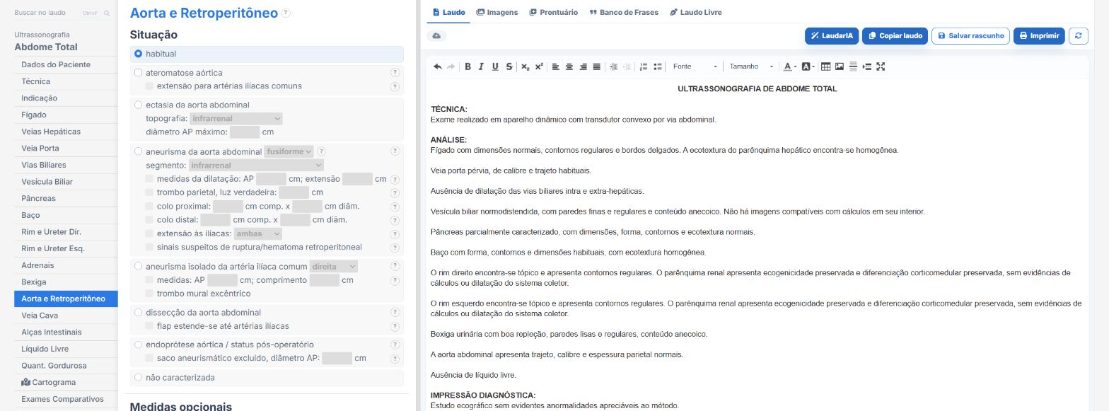
Laudário was developed to speed up report entry without losing the clinical depth that exams such as aorta ultrasound demand. Within the system, the Abdominal aorta template already comes with the structure ready to describe the suprarenal, juxtarenal and infrarenal segments, with dedicated fields for maximum AP diameter, longitudinal extent, mural thrombus characteristics, parietal calcifications and common iliac measurements, exactly the data the vascular surgeon needs to make a decision.
In practice, this means:
- A ready structure to describe each segment of the aorta, with standardized phrases adjustable to your reporting style.
- Dedicated fields for measurements in millimeters and for characterizing mural thrombus, preventing important criteria from being forgotten during fast typing.
- A standardized conclusion that can be adjusted to reflect both normal exams and aneurysm follow-up with comparison over time and an automatic suggestion of surveillance interval according to the diameter range.
- Integration with the aorta and iliac Doppler templates and the total abdomen template, making combined reports easier when the exam identifies relevant incidental findings in neighboring organs.
- Professional printing with QR Code and a digital version of the report, adding traceability and more perceived value when delivered to the patient and the requesting physician.
- Support from LaudarIA, the integrated artificial intelligence, to help build phrases, conclusions and fine adjustments to the text, particularly useful in complex descriptions such as aneurysms with heterogeneous thrombus, associated iliac findings or patients in post-EVAR follow-up.
The result is a standardized abdominal aorta report that is quick to produce and, above all, more useful to the vascular surgeon who will receive the exam. In a high-volume ultrasound practice, standardizing the aortic measurement and keeping the description of the necks, thrombus and iliac arteries always complete means less rework, greater diagnostic confidence and more value added to every exam delivered, especially in a condition in which the diameter recorded in the report is, quite literally, what defines the surgical indication.
If you are not yet familiar with it, it is worth trying: Laudário offers a 15-day free trial, no credit card required, with immediate access to all exam templates, including abdominal aorta, aorta and iliac Doppler, and the other vascular and abdominal routine protocols.
The ultrasound reporting platform built by sonologists. We write about reports, classifications and standardization from the real routine of the practice. Who writes and how.
Educational content for healthcare professionals. It does not replace clinical judgment or the guidelines adopted by your service. Editorial policy.