Renal artery Doppler: stenosis, renal-aortic ratio and intrarenal indices
Peak systolic velocity, renal-aortic ratio, the tardus parvus pattern in the intrarenal arteries and what the resistive index really tells you. How to write a report that answers whether or not there is stenosis.
 LaudárioUltrasound reports in practice
Renal artery Doppler is usually requested with one objective question: is there a renal artery stenosis that explains the hypertension or the worsening renal function? It is a long exam, difficult in obese patients or those with a lot of bowel gas, and it depends on measuring the right velocities in the right places. A report that merely says "flow present in the renal arteries" does not answer the question.
This text shows which parameters to measure, the direct and indirect criteria for stenosis and how to organize the report so that the nephrologist and the vascular surgeon find the answer.
When the exam is requested
- Hypertension with onset before age 30 or after age 55, resistant or difficult to control.
- Worsening renal function after starting an ACE inhibitor or angiotensin receptor blocker.
- Asymmetry in kidney size.
- Sudden, recurrent pulmonary edema with no obvious cardiac cause.
- Follow-up of angioplasty with stenting.
- Suspected fibromuscular dysplasia, typical of young women.
The technique
The exam has two parts, and both need to appear in the report:
- Direct assessment of the renal arteries, from the origin at the aorta to the hilum, via the anterior and lateral (translumbar) approaches. The origin is where most atherosclerotic stenoses are found; the middle and distal thirds are the typical territory of fibromuscular dysplasia.
- Indirect assessment of the intrarenal arteries (segmental or interlobar) in the upper, middle and lower poles of each kidney.
The kidney size (longitudinal diameter) and parenchymal thickness should also be recorded, because a small kidney changes the interpretation and the treatment indication. So should the presence of accessory renal arteries, which are common, may have their own stenosis and can go unnoticed.
Direct criteria for stenosis
- Peak systolic velocity (PSV) in the renal artery above 180 to 200 cm/s, depending on the criterion adopted by the service, is the main sign of hemodynamically significant stenosis (generally 60% or more).
- Renal-aortic ratio (RAR): renal artery PSV divided by the aortic PSV at the level of the renal arteries. A RAR above 3.5 reinforces significant stenosis. It is especially useful when the aorta has altered velocity, as in patients with very high or very low cardiac output.
- Post-stenotic turbulence and focal aliasing on color Doppler.
- Absence of flow in the renal artery, with a small kidney and a very dampened intrarenal signal, suggests occlusion.
The report should include the PSV of each renal artery (origin, middle and distal thirds, when obtained), the aortic PSV and the calculated RAR.
Indirect criteria: the tardus parvus pattern
When there is a significant stenosis, the flow reaching the kidney is dampened: the waveform rises slowly and with a low peak. This is the tardus parvus pattern, assessed in the intrarenal arteries:
- Acceleration time increased, above about 70 ms.
- Acceleration index reduced, below about 3 m/s².
- Loss of the early systolic peak in the waveform morphology.
Indirect criteria are useful when the origin of the artery is not well seen, but they are less sensitive for moderate stenoses. Alone, they suggest; combined with the direct ones, they confirm.
The resistive index
The intrarenal resistive index (RI) is not a criterion for stenosis. It mainly reflects the resistance of the parenchyma and small vessels:
- An RI above 0.70 to 0.80 is associated with parenchymal disease, nephrosclerosis and a poorer response to revascularization.
- A difference in RI between the two kidneys above 0.05 may suggest stenosis on the side with the lower RI.
In practice, an elevated RI in both kidneys suggests that the problem lies in the parenchyma, and this may weigh against the indication for angioplasty. It is worth recording the RI of each kidney, with the mean of the three poles.
Limitations that need to be written down
The exam is technically difficult, and not visualizing the origin of a renal artery is not the same as it being normal. The report should state clearly:
- Which segments of each artery were assessed.
- Whether accessory arteries were sought.
- The limitations (body habitus, gas, difficulty holding breath).
When the assessment is incomplete and clinical suspicion is high, a recommendation for CT angiography or MR angiography is part of a responsible report.
How to structure the report
- Kidneys: dimensions and parenchyma, on each side.
- Aorta: PSV at the level of the renal arteries.
- Right and left renal artery: PSV per segment, RAR, presence of turbulence.
- Intrarenal: RI per pole and mid-kidney, acceleration time and index.
- Accessory arteries.
- Diagnostic impression: no signs of stenosis, significant stenosis (with side and segment) or inconclusive exam.
For the rest of the renal assessment, see our text on kidney and urinary tract ultrasound.
How Laudário helps
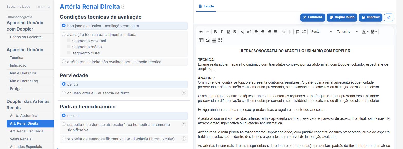
Laudário has templates for renal artery Doppler, urinary tract with Doppler and renal transplant. In practice:
- PSV fields per segment, with the renal-aortic ratio calculated automatically.
- Resistive index and acceleration time of the intrarenal arteries per pole.
- Accessory arteries and technical limitations as their own fields.
- Diagnostic impression assembled from the criteria marked.
Try it for 15 days free, no credit card required.
The ultrasound reporting platform built by sonologists. We write about reports, classifications and standardization from the real routine of the practice. Who writes and how.
Educational content for healthcare professionals. It does not replace clinical judgment or the guidelines adopted by your service. Editorial policy.