Carotid and vertebral Doppler: how to structure the report when assessing stroke risk and carotid stenosis
How to structure the carotid and vertebral Doppler report: segment-by-segment description, plaque characterization, velocities (PSV, EDV, ICA/CCA ratio), the 2003 SRU stenosis criteria and a conclusion that guides management.
 LaudárioUltrasound reports in practice
The carotid and vertebral Doppler report has to do three things unambiguously: describe each arterial segment bilaterally, quantify velocities at standardized points, and conclude with the degree of stenosis according to established criteria (NASCET, the 2003 Society of Radiologists in Ultrasound Consensus). In practice, this means recording the intima-media complex, characterizing each plaque by location, extent, thickness, echogenicity and surface, measuring the PSV and EDV of the internal carotid artery, calculating the ICA/CCA ratio and assessing the direction of flow in the vertebral arteries. The conclusion should tell the referring physician, in a few lines, whether the case calls for clinical follow-up, intensified medical therapy or vascular referral.
This demand for standardization is not perfectionism. Carotid Doppler concentrates high-impact decisions: risk stratification in asymptomatic patients, indication for antiplatelet therapy and intensive lipid-lowering treatment, evaluation for endarterectomy or stenting, and serial follow-up of plaques. Without a fixed outline, the same study receives completely different reports depending on the operator: some describe only the dominant finding, others list velocities without context, others omit the ICA/CCA ratio, the plaque characterization or the status of the vertebral arteries. In patients with a potential indication for revascularization, the difference between moderate and critical stenosis determines management.
Indications and data that should appear in the identification
The report should state the clinical indication right at the beginning: investigation of a cervical bruit, screening of a high cardiovascular risk patient (hypertension, diabetes, dyslipidemia, smoking, coronary artery disease or peripheral arterial disease), neurological symptoms (TIA, amaurosis fugax, prior stroke), preoperative cardiac surgery evaluation, follow-up of a known plaque, or post-revascularization surveillance (carotid endarterectomy or stent).
Also record the equipment and transducer used (generally a high-frequency linear transducer, with a convex or sector transducer for short necks or obese patients) and the technical limitations encountered: neck adiposity, shadowing from extensive calcifications, inability to tolerate neck hyperextension, tracheostomy, dressings. This block protects the practitioner in cases of a partial study and gives context to the referring physician.
Equally relevant are the data that influence hemodynamic interpretation: blood pressure at the time of the study, heart rate, presence of arrhythmia (especially atrial fibrillation), use of vasoactive medications and, in serial studies, the date of the prior study with an explicit comparison of changes over time. Small variations in peak systolic velocity may reflect changes in cardiac output and not necessarily progression of stenosis.
Segments to be evaluated: the backbone of the report
Segment-by-segment description is the natural backbone of the carotid report and follows the anatomy of the extracranial territory: intima-media complex, right carotid system (common, bulb, internal and external), the same sequence on the left, vertebral arteries and conclusion. A simple mention of "patent carotids" should be replaced by an explicit description of each segment studied.
- Common carotid artery (CCA): proximal, mid and distal segments, with caliber, course, intima-media thickness (IMT) measurement in the distal segment, PSV, EDV and resistive index.
- Carotid bulb: transition region with physiologically turbulent flow; preferential site of atherosclerotic plaque formation. Detail the topography (anterior, posterior, medial or lateral wall).
- Internal carotid artery (ICA): proximal, mid and distal segments accessible to ultrasound; main target of the hemodynamic analysis for stenosis stratification.
- External carotid artery (ECA): distinguished from the ICA by the presence of cervical branches, a high-resistance spectrum and the response to the "temporal tap".
- Vertebral arteries: evaluation of the V2 segment (between the transverse processes) is mandatory, with analysis of flow direction to rule out reversal (subclavian steal syndrome) and of the spectral pattern.
- Subclavian arteries: evaluation of the proximal accessible segment, especially when there is a pulse or blood pressure asymmetry between the upper limbs.
For each artery, describe the situation as normal, with a nonstenotic plaque, with a stenotic plaque and its respective degree, or with occlusion. Keeping the blocks symmetric between the two sides makes reading easier, as well as comparison with prior studies and transcription into the electronic medical record. Whenever there is doubt about the technical accessibility of a segment, document it.
How to characterize a carotid plaque in the report
The description of a carotid plaque must go well beyond "atheromatous plaque in the right bulb". Established morphological and hemodynamic criteria allow risk stratification and guide management. Each relevant plaque should be described in terms of:
- Location: segment (distal CCA, bulb, proximal/mid ICA), affected wall and laterality.
- Extent: longitudinal length in millimeters.
- Thickness: measured at the point of greatest thickness, in millimeters.
- Echogenicity: hypoechoic (predominantly lipid/hemorrhagic, classically more unstable), isoechoic (fibrolipid), hyperechoic (fibrous) or with posterior acoustic shadowing (calcified).
- Homogeneity: homogeneous or heterogeneous; heterogeneous plaques with hypoechoic areas may suggest intraplaque hemorrhage or lipid/necrotic content.
- Surface: regular, irregular or ulcerated (a niche with flow on color Doppler, more than 2 mm deep and more than 2 mm wide).
- Hemodynamic significance: estimated percentage reduction in diameter/area and correlation with the velocities obtained on the Doppler study.
Hypoechoic, heterogeneous plaques with an irregular surface, or ulcerated plaques, are considered to carry a higher embolic risk, regardless of the degree of hemodynamic stenosis. This characterization should be stated explicitly in the report, since it directly influences the indication and intensity of medical treatment.
Velocities and criteria for stenosis stratification
Hemodynamic analysis is the quantitative pillar of the carotid report. Measurements should be obtained with an appropriate angle correction (ideally ≤ 60°), with the sample volume centered on the highest-velocity jet, and in all relevant segments: distal CCA, bulb, and proximal, mid and distal ICA. The fundamental parameters include:
- ICA peak systolic velocity (PSV): the most important parameter for stenosis stratification.
- ICA end-diastolic velocity (EDV): particularly useful in severe stenoses (≥ 70%).
- ICA/CCA ratio: ICA PSV divided by the distal CCA PSV; it corrects for variations in cardiac output and is especially useful in patients with low flow or contralateral carotid disease.
- CCA PSV and spectral pattern: changes suggest upstream disease (subclavian or aortic arch stenosis) or downstream disease (distal ICA stenosis that is not accessible).
- ECA spectral pattern: useful for differentiating it from the ICA and for identifying collateral circulation.
The Society of Radiologists in Ultrasound (SRU) Consensus (2003) is the most widely adopted system for stratification:
| Degree of stenosis | ICA PSV | ICA EDV | ICA/CCA ratio | Morphological finding |
|---|---|---|---|---|
| No stenosis | < 125 cm/s | n/a | n/a | No visible plaque |
| < 50% | < 125 cm/s | n/a | n/a | Visible plaque |
| 50-69% | 125 to 230 cm/s | 40 to 100 cm/s | 2.0 to 4.0 | Plaque with luminal narrowing |
| ≥ 70% (without near-occlusion) | > 230 cm/s | > 100 cm/s | > 4.0 | Plaque with marked luminal narrowing |
| Near-occlusion | Velocities may be low or undetectable | Threadlike residual lumen; morphology and distal flow take precedence over velocity alone | ||
| Total occlusion | No flow on color, spectral and power Doppler | Pre-occlusive "thump" sign in the ipsilateral CCA | ||
The criteria must be interpreted together: no single parameter defines the stenosis. Discrepancies among PSV, EDV and the ICA/CCA ratio should be stated in the report and justified (low cardiac output, severe contralateral stenosis, tortuosity, calcifications with acoustic shadowing that limit the window).
Intima-media thickness and markers of subclinical risk
Measurement of the intima-media thickness (IMT) of the distal common carotid artery is a validated marker of subclinical atherosclerosis and overall cardiovascular risk. It should be performed on the far wall, in a plaque-free segment, about 1 cm from the bulb, with a magnified image and, ideally, with automated tools when available. Values above 0.9 mm in adults are traditionally considered increased, although the cutoff varies with age, sex and the reference adopted.
The report should record the IMT bilaterally in millimeters, indicate the measurement point, give the corresponding percentile for age and sex when available, and state whether there is diffuse or focal thickening or whether the value is within normal limits. This is key information in cardiovascular screening and in following patients on statins. The presence of plaque, by itself, is considered a stronger risk marker than IMT alone and should always be described.
Vertebral arteries: what must not be forgotten
Evaluation of the vertebral arteries is a mandatory part of the study and a frequent source of incomplete reports. The V2 segment is the most accessible and should be studied bilaterally for:
- Flow direction: antegrade (normal), retrograde (subclavian steal syndrome) or alternating (partial steal / pre-steal).
- Spectral pattern: low-resistance waveform, similar to that of the ICA; changes suggest proximal or distal stenosis.
- Peak systolic velocity: significant asymmetry between sides or very low values may indicate vertebral hypoplasia or proximal disease.
- Caliber: differences in caliber are common (hypoplasia) and should be described to avoid misinterpretation in future studies.
In patients with blood pressure asymmetry between the upper limbs, with vertebrobasilar symptoms or with clinical suspicion of subclavian disease, it is essential to document the pattern of the ipsilateral subclavian artery and the response of the vertebral artery to the reactive hyperemia maneuver.
Additional extravascular findings
Reserve a block for the extravascular findings frequently encountered during the cervical sweep: incidental thyroid nodules, lymphadenopathy, cervical cysts, salivary gland abnormalities, enlarged parathyroids. These findings should not be ignored, and the safest way to record them is in a dedicated block, with a recommendation for further evaluation when pertinent.
Conclusion and clinical guidance
The conclusion should be hierarchical and objective, summarizing in a few lines: the presence or absence of plaques and their risk features, laterality, the estimated degree of the most significant stenosis in each internal carotid artery (according to the consensus criteria adopted), the patency of the vertebral arteries, relevant extravascular findings and the suggested management: clinical follow-up, intensified medical therapy, complementary CT angiography/MR angiography or referral for vascular evaluation. Avoid repeating the entire descriptive body: the referring physician needs to read the conclusion and already know the next step.
In patients under follow-up, the conclusion must state the comparison over time explicitly: stability of the plaque and velocities, dimensional progression, change in echogenicity (potential destabilization) or increased velocities suggesting progression of the degree of stenosis.
Findings that deserve emphasis in the conclusion
- Stenosis ≥ 50% in the ICA, especially in a symptomatic patient (TIA, ipsilateral stroke, amaurosis fugax).
- Stenosis ≥ 70% in any patient, with an indication for vascular evaluation to define treatment.
- Hypoechoic, heterogeneous plaques with an irregular surface, or ulcerated plaques, even without hemodynamically significant stenosis.
- Near-occlusion or total occlusion of the ICA, with a description of the flow pattern in the CCA and in the collaterals (ECA, vertebral arteries).
- Flow reversal in a vertebral artery, suggesting subclavian steal syndrome.
- Discrepancies among PSV, EDV and the ICA/CCA ratio, justified in the report.
- Progression of the plaque or of the velocities on serial studies, even if still within the same stenosis range.
Common errors that make the report ambiguous
- Segments described collectively: terms such as "patent cervical vessels" in place of a description of each segment (CCA, bulb, ICA, ECA, vertebral and subclavian arteries) bilaterally.
- Nonstandardized IMT: measurements obtained in segments with plaque, on the near wall or without stating laterality, compromising serial comparison.
- Plaques without complete characterization: missing echogenicity, surface or estimated percentage of lumen reduction, making embolic risk stratification difficult.
- Incomplete velocities: recording only the peak systolic velocity, without EDV and without the ICA/CCA ratio, which makes it impossible to apply the stenosis criteria. Always record in cm/s, with the measurement segment and the insonation angle.
- Confusing the internal and external carotid arteries: misclassification from not using the spectral pattern and the branches as criteria, leading to serious errors in interpreting the degree of stenosis.
- Neglected vertebral arteries: a generic mention of "patent vertebral arteries", without caliber, flow direction and PSV, overlooking situations such as subclavian steal or hypoplasia.
- Conclusion disconnected from the body of the report and from the clinical picture: describing a hypoechoic plaque with an irregular surface and elevated velocities and concluding only "carotid atheromatosis", without correlating with the reason for the study.
- Ignored extravascular findings: thyroid nodules or lymphadenopathy that appear on the sweep and are not recorded, a missed opportunity for early diagnosis.
- Absolute conclusions in borderline findings: reserve "no abnormalities" for truly normal studies; in doubtful cases, describe transparently and suggest reassessment or further workup.
How Laudário helps structure the carotid Doppler report
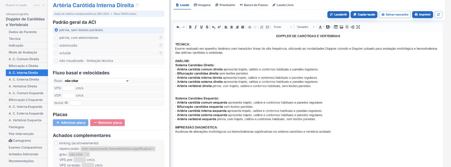
Laudário was developed to speed up report typing without losing the clinical depth that studies such as carotid Doppler demand. The Carotid and Vertebral Doppler template already comes with the complete segment-by-segment structure, organized in tabs that follow the same anatomical sequence recommended here. In practice, this means:
- Ready-made tabs for patient data, technique, intima-media complex, common carotid artery, bulb, internal carotid artery, external carotid artery and vertebral artery on each side, and additional findings.
- Standardized, customizable phrases for each segment, with options for the situation (normal, nonstenotic plaque, mild, moderate or severe stenotic plaque, near-occlusive or occlusion), echogenicity and surface.
- A specific intima-media complex block, with numeric fields for right and left IMT, ready-made phrases for diffuse or focal thickening, and integration of the measurement into the conclusion.
- Dedicated fields for velocities (PSV, EDV) and automatic calculation of the ICA/CCA ratio, preventing important criteria from being forgotten during fast typing.
- A dedicated vertebral artery block, with options for flow direction (antegrade, retrograde, bidirectional), patency, hypoplasia and PSV, making it easier to record situations such as subclavian steal.
- Automatic stenosis grading according to the 2003 SRU consensus criteria, with a conclusion that picks up the findings marked in each segment and the comparison over time in follow-up studies.
- Professional printing with a QR Code and a digital version of the report, adding traceability and more perceived value when delivering it to the patient and the referring physician.
- Support from LaudarIA, the integrated artificial intelligence, to help build phrases and fine-tune the text, particularly useful in cases with parameter discrepancies, complex plaques or extensive vascular disease.
The result is a standardized carotid Doppler report that is quick to produce and more consistent across successive studies of the same patient, which makes all the difference in following known plaques and in post-endarterectomy or post-angioplasty surveillance. Instead of starting from a blank page, you adjust an already tested template and focus on what really requires your attention: reading the images, applying the stenosis criteria and correlating with the clinical picture.
If you still build your vascular reports manually or by copying old text, it is worth a look: Laudário offers a 15-day free trial, no credit card required, with immediate access to all exam templates, including carotid and vertebral Doppler and the other routine vascular protocols.
The ultrasound reporting platform built by sonologists. We write about reports, classifications and standardization from the real routine of the practice. Who writes and how.
Educational content for healthcare professionals. It does not replace clinical judgment or the guidelines adopted by your service. Editorial policy.