Lower extremity venous Doppler: how to structure the report when DVT is suspected
The lower extremity venous Doppler report for suspected DVT should walk the deep system segment by segment, assessing compressibility, spontaneous flow, respiratory phasicity and response to distal augmentation, and close with an objective conclusion.
 LaudárioUltrasound reports in practice
The lower extremity venous Doppler report for suspected deep vein thrombosis (DVT) must answer a single question clearly: is there thrombosis or not, in which limb, in which segments and at what stage. To do so, it should walk the deep venous system segment by segment (common femoral vein, femoral vein, deep femoral vein, popliteal vein, anterior tibial, posterior tibial and peroneal veins, and the calf muscular veins), recording in each one compressibility, spontaneous flow, respiratory phasicity and response to the augmentation maneuver (distal compression). Loss of compressibility is the most specific sign of acute thrombosis; absent spontaneous flow, absent respiratory variation in the central segments and absent response to distal compression are complementary findings that reinforce the diagnosis.
DVT is a condition diagnosed essentially by ultrasound: in daily practice there is no laboratory marker or clinical finding that replaces imaging. For that reason, the way the report is built bears directly on the decision to start (or not) full anticoagulation, a treatment with bleeding risk and significant cost. A report that says only “flow present,” without detailing the compressibility of the segment, leaves the requesting physician unsure about whether thrombosis was truly excluded. A standardized report, which walks the same segments in every patient, offers diagnostic confidence, comparability on serial studies (post-treatment follow-up, suspected residual DVT), fewer requests for additional studies and more robust medicolegal documentation, especially in the emergency department, where the study is done urgently and the requesting physician usually reads only the conclusion before deciding on management.
Recommended report structure
A good structure follows the anatomy of venous return: it starts with technique and indication, records laterality, walks the deep system from the groin to the calf, evaluates the superficial system and closes with a correlated conclusion.
1. Identification, technique and indication
Record the type of study (color venous Doppler of the right, left or both lower limbs, with specific DVT assessment), the technique used (B-mode, color Doppler and pulsed Doppler; generally a high-frequency linear probe, with a convex probe in larger patients), the clinical indication (suspected DVT, edema, calf pain, post-treatment follow-up, preoperative evaluation of varicose veins) and the technical limitations: marked adiposity, significant edema, dressings, cast, pain on compression. This block gives context to the requesting physician and protects the sonographer in cases of a partial study.
2. Laterality
State explicitly which limb was examined. It may seem obvious, but it is one of the items most often missing from urgent reports, and its absence makes comparison with previous or subsequent studies of the same patient impossible.
3. Deep venous system
- Common femoral vein: evaluated in the inguinal region, near the saphenofemoral junction; compressibility, spontaneous flow, respiratory phasicity and response to distal compression.
- Femoral vein: followed along the entire thigh, from the proximal to the distal third, with serial compressions, recording any segments of noncompressibility.
- Deep femoral vein: at least in the proximal third, with compressibility and flow.
- Popliteal vein: evaluated in the popliteal fossa, supine or prone (or seated, depending on the service), with slight knee flexion, in transverse and longitudinal views.
- Anterior tibial, posterior tibial and peroneal veins: followed in pairs with the corresponding arteries, with compression and augmentation maneuvers by squeezing the calf.
- Calf muscular veins (gastrocnemius and soleal): frequently the site of clinically relevant distal DVT; they should be cited even when normal, especially in patients with symptoms localized to the calf.
For each vein, describe the status as usual, not characterized (with the reason: edema, beam attenuation, pain) or harboring deep vein thrombosis. When DVT is present, specify chronicity and caliber behavior: acute thrombus presents with a distended vein, hypoechoic content and absent compressibility; chronic thrombus, with a retracted vein, echogenic content, thickened walls and collateralized flow. Also record the extent (along its entire length or in the proximal, middle or distal third) and signs of recanalization.
4. Superficial venous system
Describe the great saphenous vein, the small saphenous vein and the superficial varicosities, recording the status (usual or with superficial thrombophlebitis), the topography of the thrombophlebitis (proximal, middle or distal third of the thigh and leg) and, when indicated, the presence and level of reflux at the saphenofemoral junction, the saphenopopliteal junction and in varicose segments. In patients complaining of varicose veins, this block is as important as the deep system.
5. Additional findings
Reserve a block for the findings that are common in patients with lower limb pain or edema: Baker cyst (intact, ruptured or complicated), muscle hematoma, collections, inguinal lymph nodes, soft tissue edema, muscle strain. They explain a large share of the suspected DVT cases that come back negative and should appear in the report, not only in a verbal comment.
6. Conclusion
The conclusion should be hierarchical and objective: presence or absence of DVT, side involved, segments affected, chronicity (acute, subacute or chronic), presence of superficial thrombophlebitis, any significant reflux and relevant additional findings. Do not repeat the whole descriptive body. The requesting physician needs to read the conclusion and already know whether the patient has an indication for anticoagulation, outpatient follow-up or alternative investigation of the complaint.
Common errors in the venous Doppler report
Even in experienced services with a defined structure, some errors recur:
- Segments not cited: concluding “no DVT” without explicitly describing the compressibility of each segment, or omitting the deep femoral, tibial and calf muscular veins: the attending physician does not know whether those veins were normal or were not evaluated.
- DVT without topography: recording “deep vein thrombosis” without indicating the vein, the third involved and the extent, which makes comparison with follow-up studies impossible.
- Undefined chronicity: failing to differentiate acute from chronic or recanalizing thrombus, leaving the attending physician without a basis to decide management and treatment duration.
- No mention of limitations: failing to record marked edema, significant pain or beam attenuation when a segment could not be well evaluated, which becomes a problem when the clinical course is poor.
- Forgotten superficial system: ignoring the great and small saphenous veins in patients with varicose veins or thrombophlebitis, requiring a new study to complete the investigation.
- Describing only the symptomatic side even when a bilateral study was requested, or citing laterality inconsistently between the header and the body of the report.
- Generic conclusion: “unremarkable” or “nonspecific abnormalities” without objectively answering the clinical question, forcing the requesting physician to read the entire report to decide management.
How Laudário helps with the DVT report
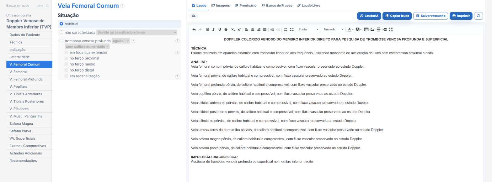
Maintaining this standardization in every study, especially on busy shifts with patients referred from the emergency department, is the great practical challenge. Laudário was designed for exactly this reality:
- Specific templates for Lower Limb Venous Doppler and Lower Limb Venous Doppler (DVT), organized in tabs (patient data, technique, laterality, common femoral vein, femoral, deep femoral, popliteal, anterior tibial, posterior tibial, peroneal, calf muscular veins, great saphenous, small saphenous, superficial veins, additional findings and conclusion).
- Ready-made fields for compressibility, spontaneous flow and response to distal compression in each segment, preventing omissions.
- Standardized, customizable phrases for each vein, with status options (usual, not characterized due to edema or attenuation, acute or chronic DVT, increased or reduced caliber, along the entire length or in the proximal, middle and distal third, recanalizing), keeping terminology consistent across successive studies of the same patient.
- A specific superficial thrombophlebitis block for the great saphenous vein, small saphenous vein and varicosities, with detailed topography in the thigh and leg.
- Automated laterality throughout the report: when you choose right, left or both limbs, the system replicates the information in the header and in the descriptive blocks.
- Associated findings (Baker cyst, muscle hematoma) described without leaving the same template.
- A conclusion integrated with the description, which picks up the findings marked in each vein, the chronicity and the additional findings, avoiding inconsistencies between the body of the report and its closing.
- Professional presentation of the final report, with the clinic header, electronic signature and a digital version with a validation QR Code, useful in urgent reports that need to circulate quickly among physicians and departments.
- A complete library covering venous and arterial Doppler of the limbs, carotids, abdomen, breast, thyroid, obstetric, morphology, gynecologic, small parts and joint studies, in a single online system.
In practice, the time spent typing segment by segment, remembering all the criteria and formatting the urgent report shrinks to a few clicks. Instead of starting from a blank page for each emergency room patient, you adjust an already tested template and focus on what really matters: careful review of the images and clinical interpretation of the findings.
If your service handles suspected DVT cases frequently, it is worth seeing Laudário in practice. Try it free for 15 days, with no credit card, and see within a few studies the difference that a system built for the ultrasound routine makes in report production.
The ultrasound reporting platform built by sonologists. We write about reports, classifications and standardization from the real routine of the practice. Who writes and how.
Educational content for healthcare professionals. It does not replace clinical judgment or the guidelines adopted by your service. Editorial policy.