Lower Limb Venous Mapping: Varicose Veins, Reflux and the CEAP Classification
Reflux testing only counts when the patient is standing: supine, incompetent valves look competent. See the maneuvers, the 0.5 and 1.0 second cutoffs, the criteria for an incompetent perforator, pelvic escape points and the CEAP that most reports lack.
 LaudárioUltrasound reports in practice
There are two lower limb venous Doppler exams, they answer opposite questions, and confusing them is the origin of most poor venous reports. One asks "is there thrombus here?": it is the DVT study, done with the patient lying down, compressing vein by vein. The other asks "is the blood flowing back?": it is venous mapping, and it only works with the patient standing.
A mapping done supine is not a bad mapping: it is an exam that does not measure what it set out to measure. Supine, the hydrostatic column that provokes reflux simply does not exist, and frankly incompetent valves look competent. This text walks through the reflux exam: the position, the maneuvers, the time cutoffs, the segments, the perforators and the CEAP classification, which is what turns the report into the surgeon's language.
The two exams, and why the report must say which one it is
The distinction is anatomic and functional at the same time:
| DVT study | Venous mapping | |
|---|---|---|
| Question | Is there thrombosis? | Is there reflux? Where does it come from? |
| Position | Supine | Standing (or steep reverse Trendelenburg) |
| Central maneuver | Transducer compression | Valsalva and distal compression |
| Organization | By vein, one by one | By function: superficial → perforators → deep |
| Urgency | High (anticoagulation today) | Elective (planning) |
The two can coexist in the same patient, and often do: post-thrombotic syndrome is exactly reflux caused by prior thrombosis. But the report must make explicit which question was answered. If you want the detail of the thrombosis study, it is in Lower limb venous Doppler: how to structure the report in suspected DVT. Here the subject is reflux.
Patient position is the datum that validates the exam
This is the most important piece of information in this text, and the one most often missing from reports: reflux testing is done standing, with weight on the contralateral limb and the examined limb relaxed, in slight external rotation. The acceptable alternative is steep reverse Trendelenburg, with the table tilted enough to reproduce the hydrostatic column.
The reason is mechanical. Reflux is the failure of the valve to prevent retrograde flow under pressure. Supine, there is no column of blood pushing downward, so the pressure the valve would need to withstand does not exist. The result is a systematic false negative: a patient with a frankly incompetent great saphenous vein can have a "normal" exam just because they were lying down.
And there is a practical consequence that almost no report records: the report should state the position in which the exam was performed. Without that, whoever receives the document cannot know whether "no reflux" means "there is no reflux" or "I did not look properly". It is the venous equivalent of not recording bowel preparation in an endometriosis mapping.
The maneuvers, and what each one tests
There is no single maneuver. Each tests one segment and has its own limitation:
- Valsalva: increases intra-abdominal pressure and tests the proximal valves: saphenofemoral junction, common femoral, high femoral segment. It is the maneuver of choice for the crossing. Its limitation is anatomic: if there is a competent valve above the point examined, the Valsalva pressure does not reach down there, and the test gives a false negative in distal segments.
- Manual distal compression (calf or thigh) followed by sudden release: reflux is sought on release. It serves any level and is the most used maneuver day to day.
- Pneumatic cuff with standardized rapid inflation and deflation: the most reproducible and the reference in studies, precisely because it eliminates the variability of the examiner's hand. Less practical in routine.
- Paraná maneuver (active contraction-relaxation): a useful alternative when the patient does not tolerate the others.
A mature report records which maneuver demonstrated the reflux, because this qualifies the finding. "Reflux detected on distal compression and on the Valsalva maneuver" is more robust than either one alone.
The time cutoffs: 0.5 and 1.0 second
Physiologic reflux exists. Every valve takes an instant to close, and a brief return is not disease. What defines incompetence is the duration of the retrograde flow, and the cutoffs differ by territory:
- Superficial veins (great saphenous, small saphenous, tributaries) and perforators: pathologic reflux above 0.5 second.
- Femoropopliteal deep veins: the cutoff is more permissive, above 1.0 second.
- Tibial and calf veins: also 0.5 second.
The asymmetry is not arbitrary: the deep system physiologically tolerates a longer return, and applying 0.5 s to the deep veins overdiagnoses deep incompetence, an error with real consequences, because deep incompetence changes the indication for superficial surgery.
In honesty: measuring this time requires the spectral tracing with the caliper on screen. Marking "sustained reflux" by eye, without timing it, is an impression dressed up as a number. If you did not measure it, the report can say "reflux evident on maneuvers", which is honest, instead of citing a cutoff that was not applied.
The map: where reflux enters and where it exits
Mapping does not list veins: it reconstructs a circuit. Every varicose territory has an escape point (where blood leaks from the deep to the superficial system) and a reentry point (where it returns). Describing both is what allows surgery to be planned.
Saphenofemoral junction and great saphenous vein
The SFJ is the most common escape point. The report should say whether it is competent and, if not, whether there is dilation of the crossing and inguinal neovascularization, the latter being the marker of postoperative recurrence.
In the great saphenous vein, record the diameters by level (crossing, mid thigh, knee, proximal leg, mid leg, malleolus) and the extent of reflux: from where to where. A saphenous vein that refluxes only in the thigh and one that refluxes down to the malleolus are different diseases, with different techniques. Diameters matter because they guide the choice between thermal ablation, foam and conventional surgery.
Pay attention to two anatomic details that change management: the saphenous compartment (the "Egyptian eye") confirms that the vessel examined is the saphenous vein and not a tributary, and a saphenous vein that leaves the compartment and becomes subdermal is a relevant technical datum for ablation. And the accessory saphenous veins (anterior and posterior) are a frequent cause of "recurrence" that is actually disease never treated.
Small saphenous vein and Giacomini
The saphenopopliteal junction is anatomically variable: it may drain high, into the gastrocnemius, or may not even exist in the typical form. Describing the variation found is essential: the surgeon needs to know where to look.
The vein of Giacomini deserves its own mention: it is the communication between the small and great saphenous veins on the posterior thigh, and can transmit reflux from one system to the other. Small saphenous reflux that actually comes from the great saphenous via Giacomini has a different treatment.
Perforators
An incompetent perforator is not every visible perforator. The criteria consolidated in the literature are two, combined:
- Diameter ≥ 3.5 mm, and
- reflux ≥ 0.5 second, with flow going from the deep to the superficial system.
And there is a third element that greatly raises relevance: the topographic association with an ulcer, active or healed. An incompetent perforator under an ulcer is a direct therapeutic target; the same perforator in a leg without a skin lesion often is not.
Always record the distance, from the plantar base in the leg and from the knee joint line in the thigh, because that is how the surgeon finds it.
CEAP: the classification missing from most reports
Here lies the greatest opportunity to improve the Brazilian venous report. CEAP is the international language of chronic venous disease, and is composed of four axes. Ultrasound answers the last three directly:
- C: Clinical (this is the physical exam, not ultrasound):
- C0: no visible signs
- C1: telangiectasias or reticular veins
- C2: varicose veins
- C3: edema
- C4: skin changes (pigmentation, eczema, lipodermatosclerosis, atrophie blanche)
- C5: healed ulcer
- C6: active ulcer
- E: Etiology: Ec congenital, Ep primary, Es secondary (post-thrombotic), En not identified.
- A: Anatomy: As superficial, Ap perforators, Ad deep, An not identified.
- P: Pathophysiology: Pr reflux, Po obstruction, Pr,o both, Pn none.
A report that already describes great saphenous reflux, an incompetent perforator and absence of thrombosis has all the inputs to write Ep, As,p, Pr, and does not write it. It is information that is already in the exam and is lost by convention.
A caveat on scope: the "C" is clinical. The sonologist can record what is seen (ulcer, skin change), but the full clinical classification belongs to the vascular specialist. The E, A and P axes, on the other hand, are essentially sonographic, and reporting them adds immediate value.
Varicose veins, telangiectasias and pelvic escape points
The description of varicose veins should say which territory they belong to: great saphenous, small saphenous, or neither. Varicose veins that do not connect to any saphenous vein have another origin, and this is where the pelvic escape points come in: inguinal, perineal, gluteal and obturator.
This is an underdiagnosed and clinically important finding, especially in multiparous women with varicose veins of the medial thigh or vulvar region that "recur" after surgery. In these cases, the recurrence was never a recurrence: the pelvic escape point was never treated, because it was never described. If suspected, the workup extends to pelvic evaluation.
The deep system in the reflux exam
Even in a mapping, the deep system must be evaluated, for two distinct reasons. First, to exclude thrombosis, acute or chronic: post-thrombotic syndrome is the main cause of secondary venous insufficiency, and the finding changes the CEAP etiology from Ep to Es. Second, to look for deep reflux, with the 1.0 s cutoff, because significant deep incompetence alters the prognosis of superficial treatment and, in some scenarios, contraindicates it.
It is also worth recording duplications (a duplicated femoral vein is common and is a classic trap: one of the channels may be thrombosed and the other patent).
Pitfalls of the venous report
- Doing the exam supine: the error that invalidates everything. Without a hydrostatic column, there is no reflux to measure.
- Not recording the position: without it, the reader does not know whether "no reflux" is a finding or an artifact of method.
- Applying 0.5 s to the deep system: overdiagnoses deep incompetence, the datum that most changes management.
- Citing "sustained reflux (> 0.5 s)" without having measured it: it is an impression dressed up as a number.
- Using Valsalva alone: a competent valve above the point examined blocks the maneuver and produces a distal false negative.
- Calling every visible perforator incompetent: the criterion is diameter ≥ 3.5 mm and reflux ≥ 0.5 s.
- Not measuring saphenous diameters by level: they are what guide the choice of technique.
- Not describing the reentry point: the circuit is left half done.
- Forgetting the accessory saphenous veins and Giacomini: a frequent source of false recurrence.
- Not looking for a pelvic escape point in medial thigh varicose veins in a multiparous woman.
- Omitting CEAP: the inputs are already in the exam; it only needs to be written.
How Laudário helps structure venous mapping
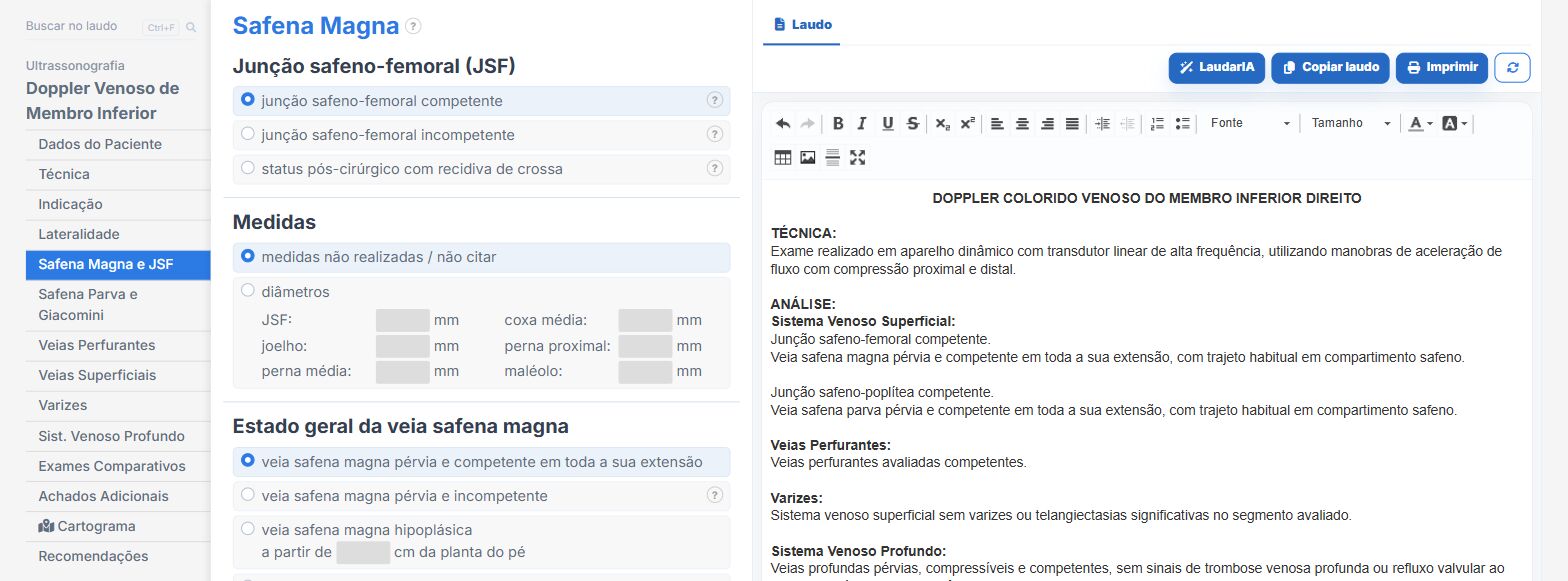
Laudário has separate modules for the two questions: Lower Limb Venous Doppler (mapping, organized by hemodynamic function) and DVT Study (organized by vein), which already avoids scope confusion at the source. In practice:
- Tabs in the order of reasoning: great saphenous vein and SFJ → small saphenous vein and Giacomini → perforators → varicose veins → deep system. The form is the circuit.
- Fields by level for the diameters of the two saphenous veins, automatically consolidated into a table in the report.
- Extent of reflux by segment, with reentry point, tributaries and draining perforator described in the same sentence.
- Perforator adder with diameter, distance (automatically referenced to the plantar base or the joint line, depending on the segment) and marking of topographic association with an ulcer.
- Pelvic escape points (inguinal, perineal, gluteal, obturator) as their own field, the finding that tends to be forgotten.
- Recording of the maneuver that demonstrated reflux in the deep system (distal compression, Valsalva or both).
- Venous cartogram with phlebogram in three views, with color coding by state (physiologic, incompetent, recanalized, occluded), exportable for printing with the report: the map the surgeon wants in hand.
- Auto-suggested recommendations according to the finding, and support from LaudarIA for multi-territory cases.
If you perform venous mapping and still build the report manually, it is worth trying: 15 days free trial, no credit card, with all vascular modules unlocked.
The ultrasound reporting platform built by sonologists. We write about reports, classifications and standardization from the real routine of the practice. Who writes and how.
Educational content for healthcare professionals. It does not replace clinical judgment or the guidelines adopted by your service. Editorial policy.