Temporal artery Doppler in giant cell arteritis: the halo sign and the report
Why ultrasound has become the first exam when giant cell arteritis is suspected, how to look for the halo sign and the compression sign, which segments to evaluate, the importance of the axillary arteries and what the report should state.
 LaudárioUltrasound reports in practice
Giant cell arteritis is the most common vasculitis above age 50 and can lead to irreversible vision loss if not treated in time. European recommendations now indicate ultrasound as the first imaging exam when clinical suspicion exists, because it is fast, accessible and able to change management on the same day. But the exam requires careful technique and a report that states clearly what was seen in each segment.
Timing matters
Wall thickening regresses with corticosteroids, sometimes within a few days. Ideally, the exam is performed before or right at the start of treatment. The report should record whether the patient is already taking corticosteroids and for how long, because a normal exam after days of treatment does not carry the same value.
Technique
- High-frequency linear transducer, with plenty of gel and minimal pressure.
- B-mode and color Doppler, adjusted for small-artery flow.
- Bilateral evaluation of the common superficial temporal artery and the frontal and parietal branches, in transverse and longitudinal planes.
- Evaluation of the axillary arteries, because large-vessel involvement is frequent and can exist even with normal temporal arteries.
The findings
- Halo sign: hypoechoic, homogeneous, concentric wall thickening, visible in both transverse and longitudinal planes. It is the most characteristic finding.
- Compression sign: when the artery is compressed with the transducer until the lumen disappears, the thickened wall remains visible. In a normal artery, the wall disappears with compression.
- Wall thickness (intima-media complex): cutoffs have been proposed for each segment, with lower values in the branches than in the trunk. Measuring and recording helps follow-up.
- Stenosis or occlusion, with increased velocity or absence of flow, are complementary findings.
Differentials and pitfalls
- Atherosclerosis thickens the wall eccentrically, irregularly and often with calcification, unlike the homogeneous, concentric halo.
- Excessive transducer pressure erases flow and distorts the wall.
- High color gain can "fill in" the halo and hide it.
What the report should include
- Corticosteroid use and duration of treatment.
- For each segment evaluated, on each side: presence or absence of halo, compression sign and wall thickness.
- Result of the axillary arteries.
- A clear conclusion: findings consistent with arteritis (with the extent) or no signs of vasculitis in the segments evaluated, remembering that a negative exam does not exclude the disease when clinical suspicion is high.
Common errors
- Evaluating only the temporal artery on one side.
- Not examining the axillary arteries.
- Omitting prior corticosteroid use.
- Describing "thickening" without saying whether it is concentric and whether it persists on compression.
- Reporting a negative result without citing which segments were evaluated.
See also our text on carotid and vertebral Doppler.
How Laudário helps
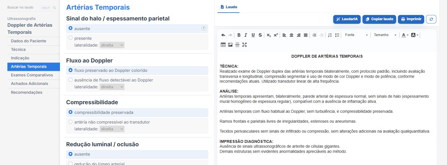
In the temporal artery Doppler template of Laudário:
- Segments on each side in their own fields, with the halo sign and the compression sign.
- Conclusion built from the findings checked.
- Recommendations in their own tab.
Try it for 15 days free, no credit card required.
The ultrasound reporting platform built by sonologists. We write about reports, classifications and standardization from the real routine of the practice. Who writes and how.
Educational content for healthcare professionals. It does not replace clinical judgment or the guidelines adopted by your service. Editorial policy.