Gallbladder ultrasound and cholelithiasis: how to describe, classify and standardize the report
How to structure the gallbladder ultrasound report: describing stones, biliary sludge, polyps and signs of cholecystitis, and correlating with the clinical picture.
 LaudárioUltrasound reports in practice
This article is part of the guide Abdominal Ultrasound: A Guide to the Findings That Actually Change Management.
Gallbladder ultrasound is probably one of the most frequently requested exams in abdominal routine, whether as a stand-alone evaluation or as part of a complete abdominal study. Although technically accessible, the way each professional describes the findings varies widely: some reports merely mention a “single stone” without dimensions, others describe the wall and contents in detail but forget the bile ducts, and many do not comment on the sonographic Murphy sign even when the clinical picture suggests it. When the report is incomplete, the surgeon and the clinician lose relevant information for deciding between expectant management, outpatient follow-up or cholecystectomy. In this article, we lay out a practical structure for the gallbladder ultrasound report, focused on cholelithiasis, biliary sludge, polyps, signs of acute cholecystitis and evaluation of the bile ducts.
Why standardize the gallbladder report?
The gallbladder is a small structure, but it concentrates an enormous amount of clinically relevant information: dimensions, wall thickness, contents, presence of mobile or impacted stones, polyps, biliary sludge, inflammatory signs and the relationship with the intra- and extrahepatic bile ducts. Without a fixed sequence, three problems appear frequently:
- Vague description of the contents: “echogenic foci with posterior acoustic shadowing” without stating the number, the size of the largest stone, mobility and location (fundus, body, infundibulum).
- Superficial evaluation of the wall: a report that only states “thin walls” or “thickened walls” without the measurement in millimeters, compromising comparison over time and risk stratification for cholecystitis.
- Polyps described without criteria: a polypoid lesion mentioned in the description without dimensions, number, base of attachment or Doppler evaluation, exactly the parameters that guide surgical management.
Standardizing the report for the gallbladder is, above all, making sure that the entire anatomy and the main differential diagnoses are always covered: dimensions, walls, contents, focal findings, sonographic Murphy sign and bile ducts, closing with a prioritized impression directly correlated with the body of the report.
Recommended structure for the gallbladder ultrasound report
A good structure for gallbladder ultrasound follows the logic of the exam: evaluate the gallbladder as a whole, describe the contents, classify focal findings, review the bile ducts and close with a clear diagnostic impression. We suggest the following blocks:
1. Identification and technique
Include the type of exam, fasting conditions (ideally six to eight hours), any technical limitations (body habitus, bowel gas distension, reduced acoustic window) and the equipment used, when relevant. This block gives context to the requesting physician and protects the professional in technically limited exams, which is especially important when fasting was inadequate and the gallbladder is partially contracted.
2. Gallbladder dimensions and walls
Describe the gallbladder in distension (volume, longitudinal and transverse diameters), contours and wall thickness measured in millimeters (usual reference value: up to 3 mm when fasting). Also comment on wall regularity, the presence of stratification, edema, a hypoechoic pericholecystic halo or adjacent free fluid. These elements support the differentiation between chronic, acute and sequelae findings.
3. Gallbladder contents
This is the most informative block of the report. For each intraluminal finding, record:
- Stones: number (single, multiple or innumerable), size of the largest stone, preferential location (fundus, body, infundibulum, neck), mobility with changes in position and presence of posterior acoustic shadowing. Stones impacted in the infundibulum or neck deserve emphasis, given their relationship with cholecystitis and Mirizzi syndrome.
- Biliary sludge: description of the pattern (homogeneous, layering, in a “ball”), relative volume and mobility. Biliary sludge has a different clinical implication from a formed stone and should be named specifically.
- Polyps: number, individual dimensions (especially the largest), location, base of attachment (sessile or pedunculated), echogenicity, and the absence or presence of mobility and of flow on color Doppler. Lesions equal to or larger than 10 mm, or with documented growth on follow-up, deserve emphasis in the conclusion.
4. Signs of acute cholecystitis
When there is clinical suspicion, explicitly describe the main sonographic signs: wall thickening greater than 3 mm with stratification, gallbladder distension, a stone impacted in the neck or infundibulum, the presence of pericholecystic fluid and a positive sonographic Murphy sign on directed compression with the transducer. In patients with acalculous cholecystitis (no stones), the remaining signs carry even more weight and should be made explicit.
5. Intra- and extrahepatic bile ducts
Even in an exam focused on the gallbladder, systematically evaluate the bile ducts: common bile duct caliber (usual reference value up to 6 to 7 mm in adults, with tolerance for post-cholecystectomy patients and the elderly), presence of intrahepatic dilatation (portal “double-barrel” sign), content of the common bile duct when visualized and the quality of the acoustic window for this evaluation. Biliary dilatation is a finding of high clinical impact and should never go unnoticed in the report.
6. Adjacent structures
Briefly comment on the adjacent liver (parenchyma and contours near the gallbladder fossa), pancreas (when visualized), subhepatic area and the presence or absence of free fluid. These data are especially useful in acute presentations and in patients in the early postoperative period.
7. Diagnostic impression and suggested management
The conclusion should be objective, prioritized and directly correlated with the descriptive body. For the gallbladder, state the main diagnosis (cholelithiasis, acute cholecystitis, scleroatrophic gallbladder, gallbladder polyps, isolated biliary sludge) and, when applicable, suggest clinical correlation, ultrasound follow-up at a defined interval or specialist surgical evaluation. The final decision always rests with the attending physician, but the report should provide all the elements to make it.
Common errors in gallbladder and cholelithiasis reports
Even with a defined structure, some recurring errors undermine the quality of gallbladder reports:
- Not stating stone dimensions: citing only “cholelithiasis” without the size of the largest stone, approximate number and location, making surgical stratification difficult.
- Confusing biliary sludge with a stone: describing mobile content without acoustic shadowing as “microlithiasis” when the correct finding is biliary sludge, entities with distinct clinical management.
- Forgetting the sonographic Murphy sign: not documenting the directed maneuver in patients with right upper quadrant pain, leaving the reader without information that has important predictive value for cholecystitis.
- Polyp described without size: mentioning a “polypoid lesion” without stating the measurement of the largest polyp, the base and the presence or absence of Doppler flow, exactly the parameters that change management.
- Omitting evaluation of the bile ducts: focusing the report on the gallbladder and not commenting on common bile duct caliber and the presence or absence of intrahepatic dilatation.
- Conclusion disconnected from the description: saying “gallbladder without abnormalities” in the impression when the body of the report describes thickened walls or biliary sludge, or vice versa.
How Laudário helps structure the gallbladder report
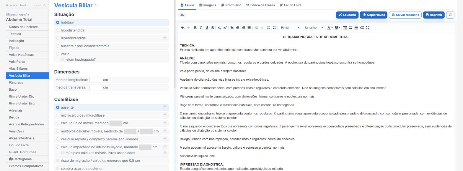
Laudário was designed for professionals who face this routine every day. For gallbladder ultrasound, and for the main abdominal protocols, the system offers:
- Ready-made, structured templates for total abdomen, upper abdomen and a focused biliary tract study, organized in the classic sequence of the exam to speed up typing.
- Standardized, customizable phrases for describing stones, biliary sludge, polyps, signs of cholecystitis and common bile duct caliber, keeping terminology consistent between reports.
- Calculations and measurements integrated into the typing flow, avoiding manual arithmetic and reducing transcription errors in dimensions and wall thickness.
- A complete library covering abdomen, pelvis, small parts, obstetrics, musculoskeletal and Doppler exams, all in a single online system, which also makes standardization across professionals in the same clinic easier.
The result is simple: less time typing, fewer forgotten findings and more consistent reports across exams of the same patient, which makes all the difference when the change in the size of a polyp, the variation in common bile duct caliber or the appearance of new inflammatory signs can change management. Instead of starting from a blank page for every patient, you adjust a template that has already been tested, keep the clinic's professional standard and focus on what really requires your attention: reading the images.
If you still put together your gallbladder reports manually or by copying old text, it is worth getting to know Laudário. Try it free for 15 days and see in practice how a well-thought-out structure saves time on every exam.
The ultrasound reporting platform built by sonologists. We write about reports, classifications and standardization from the real routine of the practice. Who writes and how.
Educational content for healthcare professionals. It does not replace clinical judgment or the guidelines adopted by your service. Editorial policy.