Gallbladder polyps on ultrasound: what to measure and when to recommend follow-up
How to tell a polyp from a stone and from biliary sludge, why morphology matters as much as size, and what the SRU and European consensus statements say about follow-up and surgery.
 LaudárioUltrasound reports in practice
This article is part of the guide Abdominal Ultrasound: A Guide to the Findings That Actually Change Management.
Gallbladder polyp is a frequent finding and, in the vast majority of cases, benign, almost always a cholesterol polyp. The problem is that the recommendation in the report tends to vary from one examiner to another: one asks for a six-month follow-up of a 3 mm polyp, another does not comment on an 11 mm one. Standardizing the description is what makes it possible to follow current consensus statements.
First: is it really a polyp?
- Stone: hyperechoic, with posterior acoustic shadowing, and moves with a change of decubitus position.
- Biliary sludge: echogenic material without shadowing, which settles in the dependent portion and shifts slowly.
- Polyp: fixed projection from the wall, without shadowing, which does not change position with decubitus.
- Adenomyomatosis: wall thickening with echogenic foci and comet-tail artifact, which should not be mistaken for a polyp.
Repositioning the patient is part of the exam. Without it, an adherent stone and tumefactive sludge become a "polyp" in the report.
What to measure and describe
- Number of polyps and the largest measurement of the largest one.
- Morphology: pedunculated (with a thin stalk or as a "ball on the wall") or sessile (broad base).
- Focal thickening of the wall adjacent to the polyp.
- Vascularity on Doppler, when assessable.
- Comparison with the prior study, with the earlier measurement.
What the consensus statements recommend
The Society of Radiologists in Ultrasound (SRU, 2022) consensus stratifies by morphology. In summary:
- Pedunculated (extremely low risk): up to 9 mm need no follow-up; from 10 to 14 mm, ultrasound surveillance; from 15 mm, surgical evaluation.
- Sessile or broad-based (low risk): up to 6 mm need no follow-up; above that, surveillance, and surgical evaluation for the larger ones.
- Focal thickening of the wall adjacent to the polyp is a higher-risk criterion and indicates surgical evaluation.
The European consensus (ESGAR and associated societies, 2022) is more conservative: it indicates cholecystectomy from 10 mm and, between 6 and 9 mm, when there are risk factors such as age over 60 years, primary sclerosing cholangitis, Asian ethnicity or a sessile base.
Because the two documents differ, a useful report gives size and morphology, which are the data that either pathway uses, and leaves management to the treating physician when the recommendation is not consensual.
Common errors
- Not changing the decubitus position and calling an adherent stone or sludge a polyp.
- Describing "small polyps" without a measurement.
- Omitting whether the polyp is pedunculated or sessile.
- Recommending follow-up for every polyp, including those the consensus statements exempt.
See also our text on gallbladder and cholelithiasis.
How Laudário helps
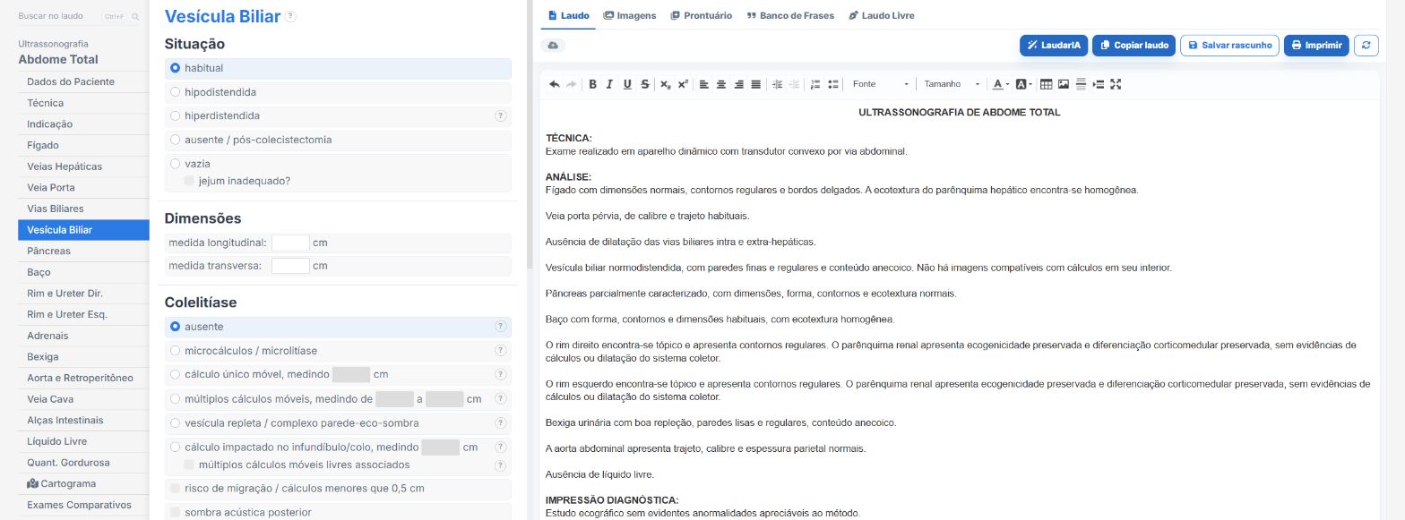
In the abdomen templates of Laudário:
- Gallbladder with stones, sludge and polyps in separate fields.
- Polyp measurement and morphology recorded for comparison.
- Follow-up recommendation on its own tab.
Try it for 15 days free, no credit card required.
The ultrasound reporting platform built by sonologists. We write about reports, classifications and standardization from the real routine of the practice. Who writes and how.
Educational content for healthcare professionals. It does not replace clinical judgment or the guidelines adopted by your service. Editorial policy.