Biliary tract on ultrasound: dilatation, common bile duct measurement and choledocholithiasis
How to measure the common bile duct, the normal limits and what changes with age and after cholecystectomy, the signs of intrahepatic dilatation, choledocholithiasis and the causes of obstruction the report should look for.
 LaudárioUltrasound reports in practice
This article is part of the guide Abdominal Ultrasound: A Guide to the Findings That Actually Change Management.
Evaluation of the biliary tract is part of every abdominal ultrasound, but it often fits in a generic line: "biliary tract not dilated". When there is jaundice, biliary pain or abnormal canalicular enzymes, that line needs to become a measurement, a location and, if there is obstruction, a probable cause. Ultrasound is the initial study; the report decides whether the workup proceeds to MRCP or to treatment.
How to measure the common bile duct
The common bile duct is measured at the porta hepatis, anterior to the portal vein, inner wall to inner wall, at the largest visible caliber. Color Doppler helps avoid confusing the common bile duct with the hepatic artery, which can cross between it and the portal vein.
Normal limits
- In adults, a common bile duct of up to about 6 mm is considered normal.
- With aging, the caliber tends to increase slightly, and somewhat larger values can be normal in the elderly.
- After cholecystectomy, the common bile duct may become larger without meaning obstruction; the finding only gains weight with intrahepatic dilatation, symptoms or laboratory abnormalities.
Therefore, the isolated measurement of the common bile duct is worth less than the whole picture: caliber, intrahepatic ducts, clinical presentation and tests.
Dilatation of the intrahepatic bile ducts
Normal intrahepatic ducts are barely seen. When dilated, they appear as tubular structures parallel to the portal branches, the so-called parallel channel sign or "double-barrel" sign. Doppler confirms that the channel without flow is the bile duct. Intrahepatic dilatation with a normal common bile duct suggests high obstruction, at the hilum; dilatation of the entire course indicates distal obstruction.
Choledocholithiasis
A stone in the common bile duct appears as a hyperechoic image with acoustic shadowing, within the duct. The sensitivity of ultrasound is limited, especially in the distal common bile duct, often obscured by duodenal gas. Therefore:
- The report should state how far the common bile duct was evaluated.
- A dilated common bile duct without a visible cause does not exclude choledocholithiasis.
- With clinical suspicion and an inconclusive ultrasound, MRCP or endoscopic ultrasound are the next steps.
Other causes of obstruction the report should look for
- Mass in the head of the pancreas, with associated pancreatic duct dilatation (double duct sign).
- Mass at the porta hepatis (Klatskin tumor).
- Lymphadenopathy at the porta hepatis.
- Choledochocele and choledochal cyst, in young patients.
- Air in the bile ducts (pneumobilia), after papillotomy or anastomosis, which should not be confused with a stone.
Common errors
- "Normal biliary tract" without a common bile duct measurement in a patient with jaundice.
- Calling a mildly enlarged common bile duct after cholecystectomy an obstruction, without other signs.
- Not stating that the distal common bile duct was not seen.
- Confusing the hepatic artery with the common bile duct by not using Doppler.
- Describing dilatation without looking for the cause in the pancreas and at the hilum.
See also our text on gallbladder and cholelithiasis and the abdominal ultrasound guide.
How Laudário helps
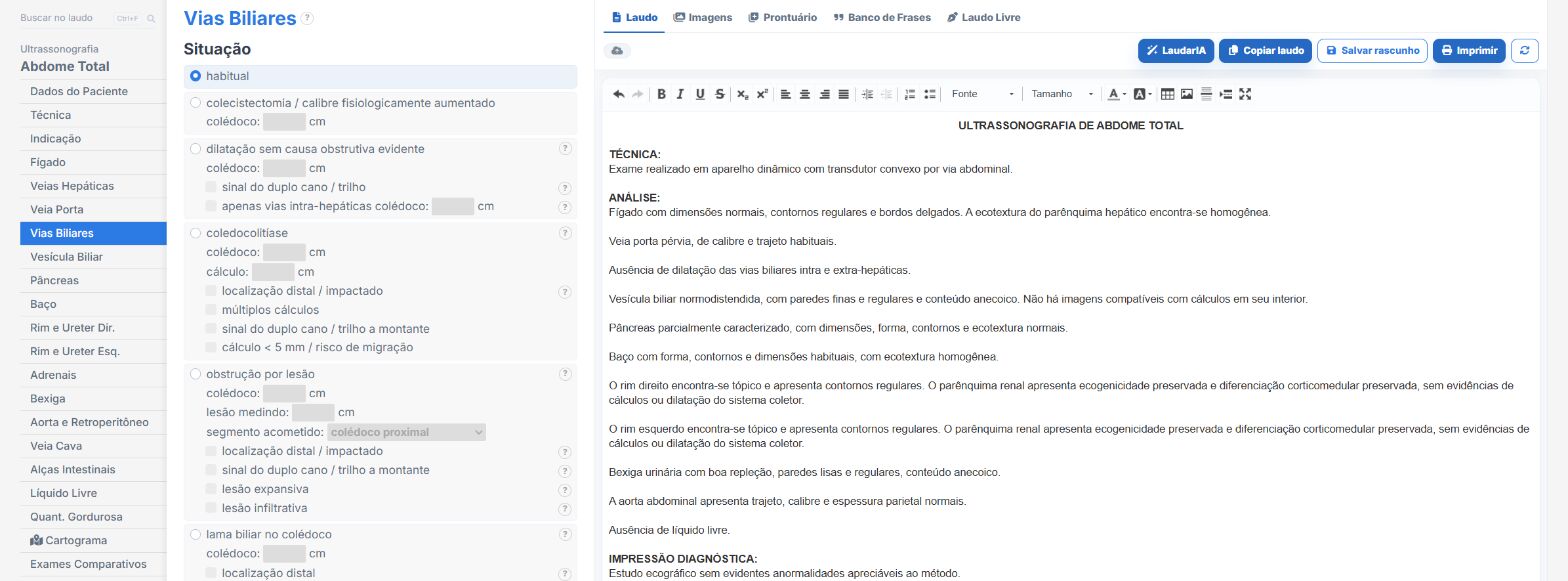
In the abdomen templates of Laudário:
- Biliary tract with common bile duct measurement and intrahepatic dilatation in their own fields.
- The conclusion is assembled from the findings, with no retyping.
- Recommendations in their own tab, such as MRCP.
Try it for 15 days free, no credit card.
The ultrasound reporting platform built by sonologists. We write about reports, classifications and standardization from the real routine of the practice. Who writes and how.
Educational content for healthcare professionals. It does not replace clinical judgment or the guidelines adopted by your service. Editorial policy.