Ultrasound report standardization: why it is essential and how to implement it in your routine
Standardizing ultrasound reports means always applying the same structure, terminology and classification criteria. The gains are diagnostic safety, consistent BI-RADS/TI-RADS/O-RADS use, legal defensibility and time.
 LaudárioUltrasound reports in practice
Standardizing ultrasound reports means always applying the same structure, the same terminology and the same classification criteria to every exam of a given type. A standardized abdominal ultrasound report, for example, always follows the same sequence of organs, describes measurements in the same unit and uses the same expressions for routine findings, such as "liver of normal size with regular contours". This does not make the report rigid or erase the physician's style: it defines a minimum structure that every exam must meet.
It is worth doing because standardization solves four problems at once: diagnostic safety (it allows comparison of serial exams of the same patient and reduces the chance of omitting descriptions in long protocols), adherence to international classifications (BI-RADS, O-RADS, TI-RADS and Bosniak only work if the criteria are applied consistently), legal defensibility (a complete description of findings, calculations and methodology is one of the best protections a physician has) and operational efficiency (less repetitive typing, faster review and integration with medical records and research). Below, each of these points and a practical path to implement standardization in your routine.
Why standardize ultrasound reports
1. Diagnostic safety
When each professional describes a finding in their own way, comparing follow-up exams becomes difficult, both for the referring physician and for any reviewer. A thyroid nodule described as "hypoechoic, with coarse calcifications" in one exam and "solid, with microcalcifications" in the next may be the same lesion or two different lesions. Standardization removes this noise. The same applies to omission: without a predefined structure, it is easy to leave out an expected item. In second-trimester anatomy ultrasound there are more than 100 morphological abnormalities that deserve systematic assessment, and a mental checklist does not always guarantee complete coverage.
2. Adherence to international classifications
Systems such as BI-RADS (breast), O-RADS (adnexal), TI-RADS (thyroid) and Bosniak (renal) only work when applied consistently. They were designed to reduce interobserver variability and guide clinical management, but they depend on the report describing exactly the criteria used to reach the category. Without standardized criteria, two sonologists may classify the same nodule differently, and this directly affects management.
3. Legal defensibility
When a report is questioned, a standardized report with a complete description of findings, calculations and the methodology used is one of the best defenses a physician can have. Descriptive variations leave room for expert witnesses to contest diagnostic criteria.
4. Operational efficiency
Standardized reports can be reviewed faster by colleagues, integrated into electronic medical records and used in clinical research. For the clinic, standardization is also a brand: the patient receives a professional document with a single visual identity, regardless of which physician performed the exam. And there is a silent cost on the opposite side. When every exam is written from scratch, losses accumulate:
- Excessive time typing information that could come ready from a template: in a routine of 20, 30 or 40 exams a day, this adds up to hours per week.
- Terminology variations among physicians on the same team, confusing the referring physician and hurting the perceived quality of the clinic.
- Inconsistency in the description of measurements, findings and conclusions.
- Difficulty training new physicians, who take weeks to understand the expected standard when it is not documented.
The pillars of a standardized report
- Fixed structure per exam: identification (type, clinical indication and technique used), description of findings and conclusion. Each modality (abdomen, obstetric, MSK, Doppler) has its own optimal structure.
- Organized description of findings: always in the same anatomical order, with measurements, echotexture and relevant relationships.
- Template phrases for frequent findings: normal exam, typical findings, usual recommendations. This reduces typing time and eliminates variations with no clinical purpose.
- Automatic calculations with interpretation: volumes, Doppler velocimetry indices, gestational age, fetal weight, percentiles, trisomy risk. When the calculation comes with the clinical interpretation, the report gains consistency.
- Integrated classifications: BI-RADS, O-RADS, TI-RADS and others should appear as part of the workflow, always in the current version, rather than as a lookup in external tables.
- Objective conclusion: it should answer the clinical question in clear language for the referring physician, cite the classification category when applicable and guide management when indicated.
How to implement it in practice
Changing the way you report on your own is hard, but the path starts with simple steps:
- List the exam types in your routine and prioritize the most frequent ones.
- Create a base template for each one, with fixed topics and variable fields.
- Define the team's preferred terminology (for example, standardize "homogeneous echotexture" instead of living with variations).
- Include ready-made phrases for common situations.
- Review the templates periodically, incorporating guideline updates such as BI-RADS, TI-RADS and the obstetric classifications.
The real challenge is keeping these templates organized and accessible during the exam. Word documents solve it only partially, and they wear out quickly: templates turn into dozens of versions, phrases get lost, classification updates take a long time to reach the routine, and each physician ends up creating a personal "fork" of the clinic's standard. The solution gaining ground among Brazilian sonologists is the use of structured platforms, which already provide ready-made templates, centralize the standard for the whole team, perform calculations with clinical interpretation and build the classifications into the reporting workflow itself.
How Laudário solves this
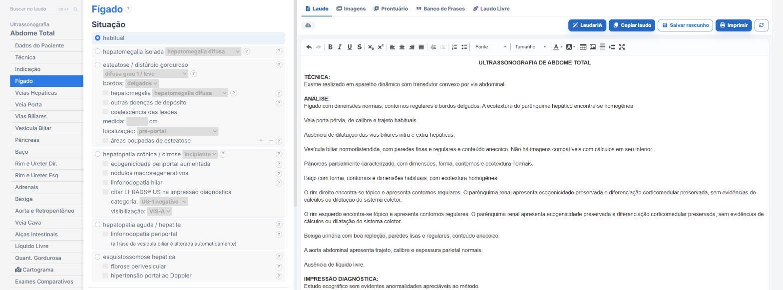
Laudário was built on exactly this principle: 72 structured templates covering abdomen, pelvis, urology, MSK, breast, gynecology, obstetrics, thyroid, Doppler and vascular, all with automatic calculations, integrated classifications (BI-RADS, O-RADS, TI-RADS, Bosniak) and phrases customizable to the physician's style. The second-trimester anatomy scan includes more than 140 morphological abnormalities already mapped, reducing the risk of omission, and LaudarIA, the built-in artificial intelligence, supports typing and speeds up report completion. Each report comes out with the clinic's visual identity, an electronic signature and an anti-fraud validation QR Code, with a verifiable digital version, ready in seconds, not minutes.
More than 671 Brazilian physicians have already produced 146 thousand reports on the platform, with reports of up to 70% less typing time per exam, without giving up quality or standardization. You can try it free for 15 days, no credit card required, at laudario.com.br.
The ultrasound reporting platform built by sonologists. We write about reports, classifications and standardization from the real routine of the practice. Who writes and how.
Educational content for healthcare professionals. It does not replace clinical judgment or the guidelines adopted by your service. Editorial policy.