Second-Trimester Anatomy Scan: How to Structure a Complete and Safe Report
How to structure the second-trimester anatomy scan report, from fetal biometry to markers, with clarity, standardization and safety.
 LaudárioUltrasound reports in practice
This article is part of the guide Obstetric ultrasound: the three-trimester guide, from dating to Doppler.
The second-trimester anatomy scan is one of the densest exams in obstetric routine: ideally performed between 20 and 24 weeks, it reviews fetal anatomy from head to toe, updates biometry, evaluates the placenta, umbilical cord and amniotic fluid, and also records markers that enter the reasoning on aneuploidy risk. A well-structured report turns this assessment into a clear clinical document, comparable throughout the pregnancy and able to guide the obstetrician's management. In this article, we organize a practical structure for the second-trimester anatomy scan report, focused on standardization, biometry, anatomy and the description of markers.
Why does the second-trimester anatomy scan require such an organized report?
Between 20 and 24 weeks, the fetus is in an ideal window for anatomic assessment: the structures are sufficiently developed, there is enough amniotic fluid to favor the acoustic window, and there is still time to confirm findings, refer to fetal medicine and plan follow-up. The report therefore has to fulfill several functions at once:
- Document the anatomic survey systematically, showing the obstetrician that every organ and system was evaluated.
- Update fetal biometry and estimate weight, comparing with reference curves and with prior exams.
- Record sonographic markers of aneuploidy when present, without inflating the risk with isolated findings.
- Evaluate adnexa: placenta, umbilical cord, amniotic fluid and cervix.
- Serve as a comparative baseline for the third-trimester obstetric ultrasound and for any reassessments.
When the report is vague or disorganized, these goals blur together: anatomy is diluted in generic phrases, biometry loses strength without comparison with curves, and markers appear without context, causing unnecessary anxiety in the pregnant patient.
Recommended structure for the second-trimester anatomy scan report
A good structure follows the logic of the exam: identify the pregnancy, record biometry, go through the anatomy in craniocaudal sequence, evaluate the adnexa and close with a clear diagnostic impression. We suggest the following blocks:
1. Identification and pregnancy data
Include the patient's name, age, last menstrual period (LMP), gestational age by LMP and by early ultrasound, number of fetuses, presentation and lie. Also record the route of the exam (transabdominal and/or transvaginal, when the cervix is evaluated) and any technical limitation: maternal body habitus, unfavorable fetal position, oligohydramnios or abdominal surgical scar.
2. Fetal biometry and estimated weight
Measure and record, at a minimum, the four classic measurements, with their respective percentiles or deviations relative to gestational age:
- BPD (biparietal diameter) and HC (head circumference): assessed in an axial plane through the thalami and cavum septi pellucidi.
- AC (abdominal circumference): axial plane at the level of the umbilical vein and stomach, without including excess soft tissue.
- FL (femur length): measuring only the ossified diaphysis, without including the epiphyses.
- Estimated fetal weight: calculated from the measurements (Hadlock or another formula used by the service), with the corresponding percentile.
Whenever prior exams exist, restate the dimensions and weight so that growth is evident. This is especially important in pregnancies with risk factors, such as hypertension, diabetes or previous growth restriction.
3. Fetal anatomy: systematic survey
Describe the anatomy in sequence, so that it is clear to the reader that the entire survey was performed. A practical order is craniocaudal:
- Head and central nervous system: cranial vault, midline, lateral ventricles (atria), thalami, posterior fossa (cerebellum and cisterna magna), cavum septi pellucidi.
- Face: orbits, lips and profile; mention the integrity of the upper lip and palate whenever possible.
- Neck: absence of masses and nuchal fold, when applicable.
- Thorax and heart: thoracic symmetry, cardiac position, rhythm, heart rate, four-chamber view, left and right ventricular outflow tracts, three-vessel and trachea view, aortic arch.
- Abdomen: stomach on the left, liver, gallbladder, kidneys (longitudinal and transverse views), bladder, umbilical cord insertion and abdominal wall.
- Spine: in longitudinal and transverse planes, evaluating alignment and skin coverage.
- Limbs: long bones, hands and feet; counting the digits is desirable when the window allows.
- Genitalia: fetal sex, when requested and technically possible, with the usual caveat about limitations.
For each item, standardize a short normal phrase in the service's template (for example, “lateral ventricles of normal dimensions”), reserving longer descriptions only for findings that depart from the expected.
4. Sonographic markers of aneuploidy
The second trimester is the phase in which several markers become visible. They should be actively sought and described clearly, without alarmism:
- Thickened nuchal fold;
- Intracardiac echogenic focus;
- Echogenic bowel;
- Mild ventriculomegaly;
- Renal pyelectasis;
- Shortened femur and/or humerus;
- Absent or hypoplastic nasal bone.
Markers must be interpreted together with clinical history, biochemical screening and prior exams. The report needs to record the finding objectively, without listing speculative diagnoses in the body of the exam: integration with the final risk is up to the obstetrician or fetal medicine.
5. Adnexa: placenta, cord, amniotic fluid and cervix
Evaluate and describe:
- Placenta: location (anterior wall, posterior, fundal, lateral), relationship to the internal cervical os, Grannum grade (when applicable) and general appearance.
- Umbilical cord: number of vessels (ideally three), placental and abdominal insertion, presence of nuchal cord when relevant.
- Amniotic fluid: subjective assessment and/or amniotic fluid index (AFI) or deepest vertical pocket, according to the service protocol.
- Cervix: length measurement by the transvaginal route in patients with an indication or per routine, with description of the internal os.
6. Doppler, when indicated
In high-risk pregnancies or those with findings that justify it, integrate the Doppler study into the report: uterine arteries, umbilical artery and, occasionally, middle cerebral artery and ductus venosus. Even when it is not part of the anatomy scan routine, record at a minimum that the study was not performed or was reserved for a dedicated exam.
7. Diagnostic impression and suggested management
The conclusion should be objective and correlated with the body of the report. In general, it covers:
- Single intrauterine pregnancy, live, with sonographic gestational age;
- Fetal biometry consistent or discordant with gestational age;
- Fetal anatomy studied, with no evident sonographic abnormalities or with specific findings;
- Markers present or absent;
- Evaluation of the adnexa;
- Suggested management, when applicable (reassessment at a given interval, referral to fetal medicine, fetal echocardiography, etc.).
Common errors in the second-trimester anatomy scan report
Even with a defined structure, some recurring errors undermine report quality:
- Anatomy in a single generic sentence: “fetal anatomy without abnormalities” without detailing systems reduces the documentary value of the exam.
- Biometry without percentiles: measurements listed without comparison with reference curves make clinical interpretation difficult.
- Markers described in the middle of the anatomy: diluting an echogenic focus or pyelectasis in long sentences removes clarity from the finding.
- Conclusion disconnected from the body: a diagnostic impression that does not mention a finding described in the text, or vice versa.
- Poor comparisons: not restating weight and biometry from prior exams in pregnancies at risk of restriction or macrosomia.
- Unrecorded limitations: failing to mention when a structure could not be evaluated (fetal position, maternal body habitus) exposes the professional and hampers follow-up.
How Laudário helps structure second-trimester anatomy scan reports
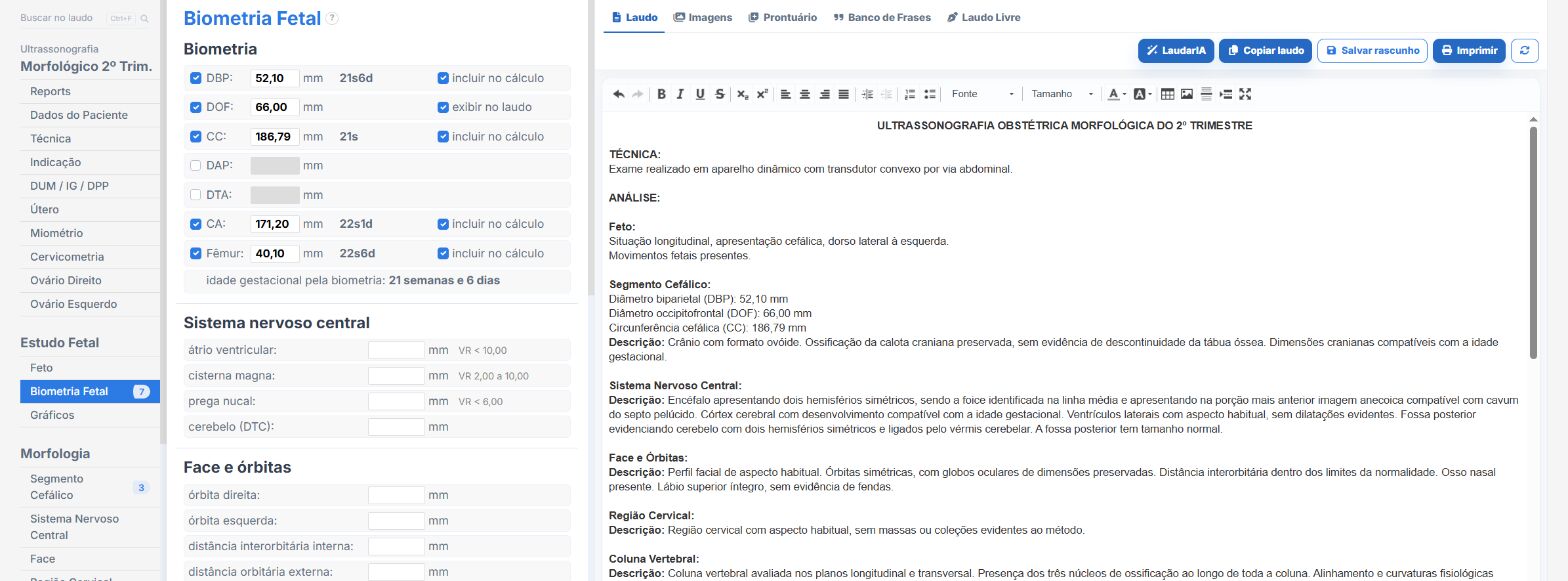
Laudário was designed for professionals who face this routine every day. For the second-trimester anatomy scan, and for the main obstetric protocols, the system offers:
- Ready-made, structured templates for first- and second-trimester anatomy scans, third-trimester obstetric ultrasound, obstetric Doppler and cervical length, organized in scanning order to speed up typing.
- Standardized, customizable phrases for fetal anatomy by system (CNS, face, thorax, heart, abdomen, spine, limbs), with short normal versions and descriptive versions for findings.
- Automatic calculations integrated into the typing workflow, including sonographic gestational age, estimated fetal weight, percentiles, AFI and cervical length measurements, avoiding manual math and reducing transcription errors.
- Comparison with prior exams, restating dimensions and fetal weight from previous reports to record growth consistently.
- A complete library covering obstetrics, breast, thyroid, abdomen, pelvis and Doppler exams, all in a single online system.
The result is simple: less time typing, fewer forgotten findings and more consistent reports across exams of the same pregnant patient, which makes all the difference in a routine where follow-up is central to management. Instead of starting from a blank page with each patient, you adjust an already tested template, keep the clinic's professional standard and focus on what really demands your attention: reading the images.
If you still build your anatomy scan reports manually or by copying old texts, Laudário is worth getting to know. Try it free for 15 days and see in practice how a well-designed structure saves time on every exam.
The ultrasound reporting platform built by sonologists. We write about reports, classifications and standardization from the real routine of the practice. Who writes and how.
Educational content for healthcare professionals. It does not replace clinical judgment or the guidelines adopted by your service. Editorial policy.