Obstetric ultrasound: the three-trimester guide, from dating to Doppler
Each trimester answers a different question, and the report needs to know which one. See the complete sequence (dating, chorionicity, nuchal translucency, anatomy scan, cervical length, growth and fetal wellbeing) and the right window for each parameter.
 LaudárioUltrasound reports in practice
Obstetric ultrasound is the only routine study in which the protocol changes completely depending on the week of gestation. The same transducer, on the same patient, answers different questions at 8, 12, 22 and 34 weeks, and a report that does not know which question it is answering at that moment ends up answering the wrong one.
This guide is the map of that sequence: what each trimester decides, which is the right study in each window, and where the pitfalls that most often generate false negatives lie. Each study has a dedicated, in-depth article, linked throughout the text.
The rule that organizes everything: dating is the foundation
Before any discussion of biometry, morphology or Doppler, there is one fact that underpins everything else: almost every obstetric parameter is relative to gestational age. Weight percentile, z-score of a cardiac structure, cervical length, amniotic fluid index, all Doppler indices: none of it means anything without the correct GA.
And the correct GA is established early, and only once. The first-trimester crown-rump length (CRL) is the most accurate parameter available for dating a pregnancy, with a margin of a few days. After the 14th week, biological variability grows and biometry loses dating power: from then on it measures growth, not age.
The practical consequence is the most important rule of obstetric imaging: once dated by CRL in the first trimester, the GA is never changed again. Redating in the third trimester because "the measurements are smaller" does not correct the date: it erases the diagnosis. A growth-restricted fetus is exactly a fetus whose measurements are smaller than its true GA; redating turns the finding you needed to see into a normal study. It is an error that happens, and it is costly.
All of this is detailed in First-trimester obstetric ultrasound: from dating to nuchal translucency.
First trimester: date, locate and screen
The first-trimester study answers four questions, in this order of urgency:
- Is the pregnancy intrauterine? This is the question that can save a life the same day. No other takes precedence.
- Is it viable? Heartbeat, and the criteria for pregnancy failure, which are deliberately conservative, because a wrong diagnosis of nonviability is irreversible.
- How many fetuses, and with what chorionicity? This is the most underrated finding of the trimester. Chorionicity defines the risk of the entire pregnancy, and it is much easier to determine between 11 and 14 weeks than later: the lambda sign and the T sign become progressively ambiguous. A monochorionic twin pregnancy not identified early is a high-risk pregnancy being managed as if it were low risk.
- What is the risk of aneuploidy? This is where nuchal translucency comes in, measured between 11 weeks and 13 weeks 6 days, with strict technical criteria: midsagittal section, neutral fetal position, adequate magnification, on-to-on calipers.
A point worth highlighting because it is frequently forgotten: an increased NT (≥ 3.5 mm) is an indication for fetal echocardiography even with a normal karyotype. The association with congenital heart disease is independent of chromosomal abnormality, and this is one of the most underused indications in all of prenatal care.
Second trimester: the anatomy scan
The second-trimester anatomy scan, between 20 and 24 weeks, is the longest and densest study in obstetrics. It is the complete anatomic survey, and it is where most structural malformations are detected.
It is also the study with the highest risk of false negatives from routine omission: when the protocol is long, it is easy to skip a plane on a busy day, and the report goes out declaring normal something that was not looked at. The defense is structural: the report template must be the exam sequence, so that only what was seen is stated.
Two blocks deserve special attention within it:
- Cervical length, ideally measured in the same study. It is cheap, quick and the best single predictor of spontaneous preterm birth, and an intervention exists. A complete anatomy scan that did not measure the cervix missed the chance to identify the patient who would benefit from progesterone or cerclage. Details in Cervical length in obstetric ultrasound.
- The heart, which is the most common malformation and the one that most often escapes. And here is the most important lesson of cardiac screening: the four-chamber view alone detects fewer than half of heart defects. Transposition of the great arteries, tetralogy of Fallot and truncus arteriosus have normal four chambers. Adding the outflow tracts and the three-vessel view raises detection to the range of 65% to 80%, and it costs no equipment at all, only a protocol.
The complete article on the study is at Second-trimester anatomy scan: how to structure a complete and safe report. And the cardiac deep dive (sequential segmental analysis, z-scores, Tei index and Huhta score) is at Screening fetal echocardiography: from situs to outflow tracts, with z-scores.
Third trimester: growth, wellbeing and hemodynamics
In the third trimester the question changes from "how is this fetus formed?" to "how is this fetus doing?". The study becomes functional:
- Growth: estimated weight and percentile for the GA established in the first trimester. This is where the temptation to redate appears, and it is where it must be resisted.
- Amniotic fluid: by deepest vertical pocket or AFI, with the caveat that the two methods are not interchangeable and the service should choose one.
- Doppler: umbilical artery, middle cerebral artery and ductus venosus, which together tell the story of fetal adaptation to placental insufficiency, in a reasonably predictable sequence of deterioration.
- Fetal wellbeing: when in doubt, the fetal biophysical profile, whose parameters disappear in the reverse order of nervous system maturation: tone is the last to be lost, and therefore the most serious.
The complete report for the trimester is at Third-trimester obstetric ultrasound: wellbeing, amniotic fluid and Doppler.
The right window for each thing
Perhaps the most useful piece of information in this guide: almost every obstetric parameter has a window in which it is reliable, and outside it becomes noise.
| To answer | Ideal window | Why |
|---|---|---|
| Dating | up to 13w6d (CRL) | After that, biological variability destroys accuracy |
| Chorionicity | 11 to 14 weeks | Lambda and T signs become ambiguous as pregnancy advances |
| Nuchal translucency | 11w to 13w6d | Validity window of the parameter itself |
| Structural morphology | 20 to 24 weeks | Organs formed and still with a good acoustic window |
| Cervical length | 20 to 24 weeks | Where the predictive value for prematurity is highest |
| Fetal echocardiography | 24 to 28 weeks | Best balance between heart size and window |
| Growth and Doppler | from 26 to 28 weeks | This is when placental insufficiency manifests |
The errors that appear most often
- Redating in the third trimester. It erases the diagnosis of growth restriction instead of correcting the date.
- Not determining chorionicity early in twins. After 14 weeks it becomes much harder, and the risk of the pregnancy depends on it.
- Concluding on the heart from the four chambers. This is the source of most of the serious false negatives in screening.
- Not measuring the cervix at the anatomy scan. It loses the only chance to identify prematurity risk while an intervention still exists.
- Ignoring increased NT with a normal karyotype. It remains an indication for fetal echocardiography.
- Declaring a plane normal without having visualized it. The report should record the limitation, not presume.
- Mixing amniotic fluid methods between studies in the same follow-up.
Note that almost all of these are protocol errors, not knowledge errors. No one fails to know that transposition of the great arteries exists: they just do not look at the outflow tracts that day. That is why report structure matters so much in obstetrics: it is what turns knowledge into routine.
How Laudário helps
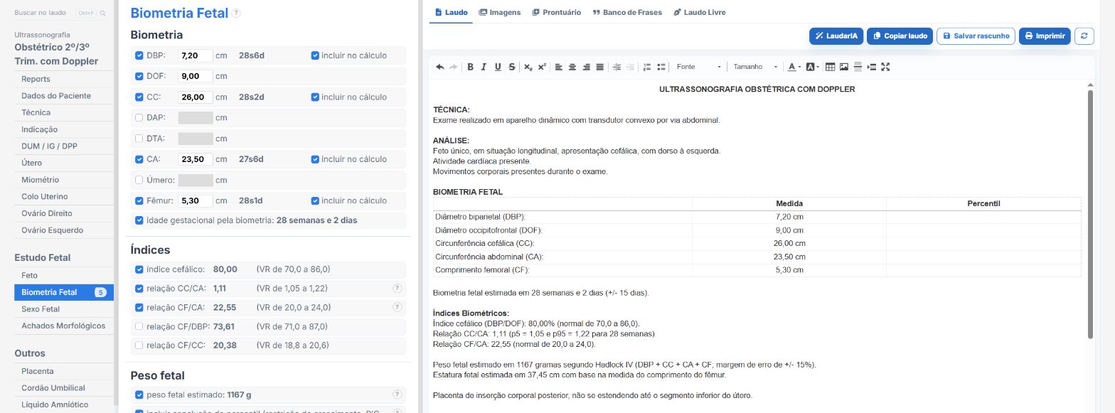
Laudário covers the entire obstetric sequence (first trimester, including twins; first-, second- and third-trimester anatomy scans; cervical length; biophysical profile; and fetal Doppler echocardiography) with modules built in scan order. In practice:
- GA derived from the recorded dating, propagating to all parameters that depend on it (percentiles, z-scores and indices) without manual recalculation.
- Z-score engine for fetal cardiac structures with GA interpolation and the reference range next to each field.
- Tab sequence that mirrors the segmental sequence, so that the form guides the study and nothing is stated without having been looked at.
- Automatic calculations: estimated weight and percentile, AFI, Doppler indices, Huhta score, Tei index.
- Ready-made heart disease templates for the most frequent diagnoses, to review instead of typing from scratch.
- Support from LaudarIA for complex cases and pregnancies under follow-up.
If you do obstetrics and build your reports manually, it is worth trying: 15-day free trial, no credit card, with all obstetric templates unlocked.
The ultrasound reporting platform built by sonologists. We write about reports, classifications and standardization from the real routine of the practice. Who writes and how.
Educational content for healthcare professionals. It does not replace clinical judgment or the guidelines adopted by your service. Editorial policy.