Third-trimester obstetric ultrasound: how to structure the report for fetal well-being, amniotic fluid and Doppler
Third-trimester obstetric ultrasound is one of the most sensitive studies in routine obstetrics and gynecology: the fetus is fully formed, the findings directly guide the prenatal care provider and any omission can have an immediate clinical outcome. Even so, it is also one of the reports that suffers most ...
 LaudárioUltrasound reports in practice
This article is part of the guide Obstetric ultrasound: the three-trimester guide, from dating to Doppler.
Third-trimester obstetric ultrasound is one of the most sensitive studies in routine obstetrics and gynecology: the fetus is fully formed, the findings directly guide the prenatal care provider and any omission can have an immediate clinical outcome. Even so, it is also one of the reports that suffers most from variations in structure between examiners: biometry without percentiles, no assessment of amniotic fluid, an incomplete biophysical profile and generically described Doppler velocimetry are recurring problems. In this article, we organize a practical structure for the third-trimester obstetric ultrasound report, focused on fetal well-being, amniotic fluid and Doppler.
Why does the third-trimester report require so much standardization?
Unlike the first trimester, where the focus is dating and nuchal translucency, and the second-trimester anatomy scan, which evaluates detailed anatomy, the third-trimester study is essentially a functional study. The referring physician wants to know whether the fetus is well, whether it is growing as expected, whether the placenta is doing its job and whether the amniotic fluid is adequate.
A lack of structure in the report usually causes three types of problems:
- Biometry without context: measurements in millimeters without estimated fetal weight, without percentile and without comparison with previous studies.
- Inconsistent amniotic fluid assessment: alternating use of AFI and deepest vertical pocket, without a clear criterion for which reference was adopted.
- Vaguely described Doppler: “preserved placental flow” without citing the umbilical and middle cerebral arteries, without indices and without the cerebroplacental ratio when indicated.
Recommended structure for the third-trimester obstetric ultrasound report
The structure needs to reflect the logic of the study: confirm fetal presentation, measure and estimate weight, assess the placenta and amniotic fluid, evaluate well-being with the biophysical profile and, when indicated, complement with Doppler velocimetry. We suggest the following blocks:
1. Identification and clinical data
Include maternal age, last menstrual period or gestational age from a prior dating study, number of fetuses, previous comparison study and any relevant clinical information: gestational diabetes, hypertension, prior growth restriction, high-risk pregnancies. This block gives context to everything that follows.
2. Fetal presentation, lie and activity
Describe presentation (cephalic, breech, shoulder), lie (longitudinal, transverse, oblique), fetal spine position and activity: body movements, breathing movements and tone. These data already make up three of the four ultrasound components of the fetal biophysical profile (BPP).
3. Biometry and estimated fetal weight
Present the key measurements with standardized units:
- BPD (biparietal diameter) and HC (head circumference);
- AC (abdominal circumference), measured in the correct transverse plane, through the portal sinus and fetal stomach;
- FL (femur length);
- Sonographic gestational age calculated from the measurements, ideally Hadlock;
- Estimated fetal weight in grams, with the corresponding percentile for gestational age;
- Comparison with previous studies when available: change in percentile over time matters more than an isolated percentile.
Whenever there is a discrepancy between clinical and sonographic gestational age, clarify the reference adopted for calculating weight and percentile. Redating in the third trimester is a serious error: the correct dating was done in the first trimester and should be preserved.
4. Placenta, umbilical cord and amniotic fluid
Describe placental location (anterior, posterior, lateral, fundal wall), relationship with the internal cervical os (especially in patients with placenta previa or low-lying placenta on prior studies), grade of maturity when relevant, and the presence of suspicious areas: prominent venous lakes, abruption, extensive infarcts.
For the umbilical cord, report the number of vessels (three vessels: two arteries and one vein) and, when applicable, placental insertion. Amniotic fluid can be quantified by AFI (amniotic fluid index) or by the deepest vertical pocket; the important thing is to keep a single criterion throughout follow-up of the same pregnancy and classify it as oligohydramnios, normal or polyhydramnios according to the cutoffs adopted by the practice.
5. Fetal biophysical profile and well-being
When requested, the BPP should be reported in a structured way, with a score for each of the five parameters (breathing movements, body movements, tone, amniotic fluid and cardiotocography, when available), totaling up to 10 points. Even when a formal BPP is not the goal of the study, fetal well-being should be commented on: active fetus, large movements, tone present, rhythmic heartbeat with mean rate reported in bpm.
6. Doppler velocimetry (when indicated)
In high-risk pregnancies (growth restriction, preeclampsia, diabetes, prolonged pregnancy), Doppler velocimetry is an essential part of the report. Include:
- Umbilical artery: pulsatility index (PI), resistance index (RI) and systolic/diastolic ratio (S/D), classified as normal, absent end-diastolic flow or reversed end-diastolic flow;
- Middle cerebral artery: PI and peak systolic velocity (when fetal anemia is suspected);
- Cerebroplacental ratio (CPR): calculated as the MCA PI divided by the umbilical PI, interpreted in the clinical context;
- Uterine arteries: mean PI and presence or absence of early diastolic notch, mainly in preeclampsia screening;
- Ductus venosus: evaluated in cases of severe growth restriction, paying attention to the “a” wave.
7. Diagnostic impression and suggested management
The impression should objectively include: sonographic gestational age, estimated fetal weight and percentile, amniotic fluid classification, placental status, Doppler findings and any relevant abnormality that deserves clinical emphasis: growth restriction, macrosomia, oligohydramnios, polyhydramnios, flow abnormalities. Final management always rests with the prenatal care provider, but the report should offer every element needed for it to be safe.
Common errors in the third-trimester obstetric report
Even with a defined structure, some errors repeat frequently:
- Fetal weight without percentile: reporting only the weight in grams, without correlating it with gestational age, makes it harder to diagnose growth restriction or macrosomia.
- Alternating between AFI and deepest vertical pocket: without a clear criterion, serial interpretation is compromised.
- “Normal Doppler” without numbers: in high-risk pregnancies, the prenatal care provider needs the indices, not just the conclusion.
- Forgetting the comparison: the third trimester is follow-up; without comparing with previous studies, the growth curve is lost.
- Redating gestational age from the current study: GA should follow the initial dating (first trimester), and the current study evaluates whether the fetus is growing according to that reference.
- Generic impression: “single live fetus in cephalic presentation” without citing weight, percentile, fluid and Doppler loses clinical value.
How Laudário helps structure third-trimester obstetric reports
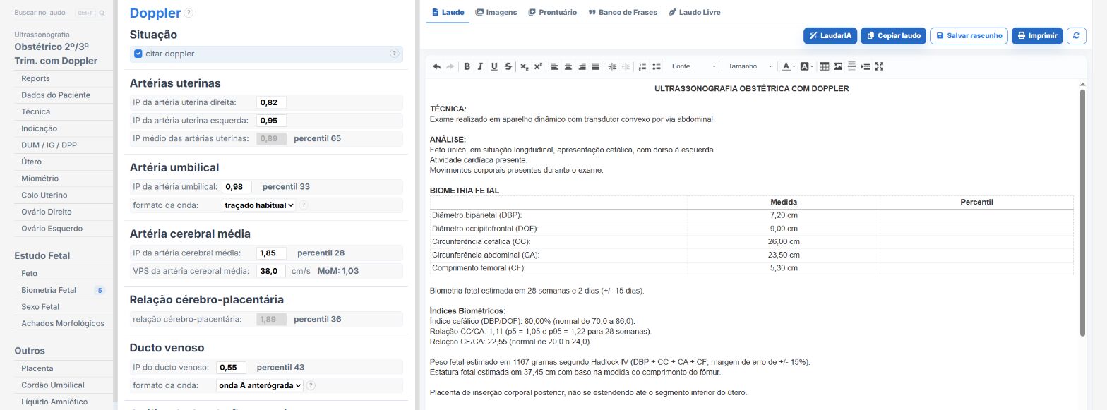
Laudário was designed precisely to make this routine more predictable, safe and fast. For third-trimester obstetric ultrasound, and for all obstetric studies, the system offers:
- Ready-made, structured templates for first-, second- and third-trimester obstetrics, anatomy scan, BPP and Doppler velocimetry, organized in the clinical sequence of the study.
- Integrated automatic calculations of gestational age, estimated fetal weight, percentile, AFI and Doppler indices, eliminating manual math and reducing transcription errors.
- Standardized, customizable phrases for presentation, well-being, placenta, cord, amniotic fluid and Doppler, keeping terminology consistent across reports for the same patient.
- Easier comparison with previous studies, helping to track the growth curve throughout the pregnancy.
- Complete library covering obstetrics, gynecology, breast, thyroid, abdomen, prostate, kidneys, urinary tract and Doppler, all in a single online system.
The result is straightforward: less typing time, fewer forgotten findings and more consistent reports across studies of the same pregnant patient, which makes all the difference in a specialty where every millimeter and every percentile can change management. Instead of starting from a blank page with every patient, you adjust a template that has already been tested, maintain the clinic’s professional standard and focus on what truly requires your attention: reading the images and caring for the pregnant patient.
If you still build your third-trimester obstetric reports manually or by copying old text, it is worth trying Laudário. Try it free for 15 days and see in practice how a well-designed structure saves time in every study.
The ultrasound reporting platform built by sonologists. We write about reports, classifications and standardization from the real routine of the practice. Who writes and how.
Educational content for healthcare professionals. It does not replace clinical judgment or the guidelines adopted by your service. Editorial policy.