First-trimester obstetric ultrasound: how to structure the report from dating to nuchal translucency
The first-trimester obstetric ultrasound report should include identification and technique, number of fetuses with chorionicity, CRL dating, viability with fetal heart rate, nuchal translucency and markers between 11 and 13 weeks 6 days, early morphology, and an objective conclusion.
 LaudárioUltrasound reports in practice
This article is part of the guide Obstetric ultrasound: the three-trimester guide, from dating to Doppler.
The first-trimester obstetric ultrasound report must follow a minimum, non-negotiable outline: identification and technique, number of gestational sacs and embryos (with chorionicity and amnionicity in multiple pregnancies), dating by crown-rump length (CRL) with the corresponding gestational age and the estimated due date, viability with fetal heart rate in bpm, nuchal translucency and other chromosomal abnormality markers when the exam is performed between 11 weeks and 13 weeks 6 days, early morphology and adnexa, and an objective conclusion that restates viability, number of fetuses, the gestational age adopted, and the estimated risk. If any of these blocks is missing, the exam loses value for pregnancy follow-up.
The reason is that the first trimester concentrates decisions that are not repeated: this is when the location of the pregnancy is confirmed, when the pregnancy is dated with the greatest precision of the entire gestation, when chorionicity is determined with confidence, and when the window for early screening for aneuploidy and preeclampsia opens. When the report is disorganized, omits measurements, or presents a conclusion inconsistent with the body of the exam, all of prenatal care is compromised. Below is a practical structure for the report, aligned with the recommendations of the Fetal Medicine Foundation (FMF) and ISUOG, focused on dating, viability, nuchal translucency, and phrase standardization.
Why does the first-trimester report require such a careful structure?
Every central point of the exam depends on objective measurements and a precise description of the findings. When the report structure fails, errors tend to repeat on three fronts:
- Inconsistent dating: gestational age by LMP and by CRL are presented without a clear criterion, or the estimated due date is not reported in a standardized way.
- Missing or poorly described markers: nuchal translucency without adequate technique, nasal bone or ductus venosus cited generically, fetal heart rate without an objective value.
- Conclusion disconnected from the body of the report: the diagnostic impression does not restate gestational age, viability, and estimated risk, leaving the treating physician without clear information for the next step.
Structuring the report does not make it rigid. It ensures that the minimum outline is always followed, so that each exam is immediately comparable with later ones and useful to the physician managing the pregnancy.
Recommended structure for the first-trimester obstetric ultrasound report
A good structure follows, in practice, the physiological sequence of early pregnancy: it starts with identification and indication, describes the gestational sac and its embryonic appendages, individualizes the embryo or fetus, records the chromosomal abnormality markers when indicated, and closes with a correlated conclusion.
1. Identification, indication, and technique
Every report should begin with complete identification: patient name, date of birth, exam date, referring physician, clinical indication (routine prenatal care, bleeding, pelvic pain, early pregnancy follow-up, chromosomal abnormality screening), and the last menstrual period (LMP) when reported. Include the type of exam (transabdominal, transvaginal, or both) and any technical limitation observed, such as body habitus, bowel loops, uterine retroversion, or unfavorable fetal position. This block gives context to the requesting physician, allows comparison of menstrual and ultrasound gestational age, justifies any limitations in assessing the markers, and protects the examiner in cases of partial exams.
2. Dating: LMP, CRL, and estimated due date
Present the gestational age by last menstrual period (when reliable) and the gestational age by ultrasound, preferably calculated by the CRL between 7 and 14 weeks. The report should give the value in millimeters and the corresponding gestational age in weeks and days, ideally with the associated margin of error. First-trimester ultrasound dating prevails over LMP when the difference exceeds 5 to 7 days. When there is a discrepancy, make clear which reference is being adopted for the estimated due date, following current recommendations. Also state the method used and, whenever possible, the reference table adopted.
3. Gestational sac, yolk sac, and location
Describe how many gestational sacs and how many embryos were identified, using objective criteria:
- Location: intrauterine or atypical location, with careful assessment for ectopic pregnancy and cesarean scar pregnancy.
- Morphology: regular or irregular contours, collapsed appearance, presence of subchorionic hematoma.
- Uterine position: fundal, anterior/posterior corpus, isthmic, with special attention to proximity to the previous scar in patients with a prior cesarean.
- Mean sac diameter (MSD): measured in three planes when indicated, with the corresponding gestational age.
- Yolk sac: presence, dimensions, and morphology.
In situations of diagnostic uncertainty (early pregnancy, miscarriage, anembryonic pregnancy, ectopic pregnancy), adopt objective viability criteria according to established references, avoiding premature conclusions.
4. Embryo/fetus: CRL, fetal heart activity, and viability
Document the presence of fetal cardiac activity and the heart rate in bpm, in addition to the CRL with the corresponding gestational age and the impression about viability. The absence of cardiac activity in an embryo with a CRL ≥ 7 mm is a criterion for diagnosing embryonic demise, according to current guidelines. In multiple pregnancies, identify each fetus individually, record chorionicity and amnionicity (lambda and T signs), information that can only be determined with confidence in the first trimester, and apply the same parameters to each one, avoiding grouped descriptions that make follow-up difficult.
5. Nuchal translucency and chromosomal abnormality markers
When the exam is performed between 11 weeks and 13 weeks 6 days (CRL between 45 and 84 mm), include the first-trimester marker block, recording adequate technique and the measurement in millimeters:
- Nuchal translucency (NT): measured in a sagittal view, with the fetus in a neutral position, adequate magnification, and calipers correctly placed at the inner borders of the translucency.
- Nasal bone: present, absent, or hypoplastic.
- Ductus venosus: A-wave positive, absent, or reversed.
- Tricuspid regurgitation: present or absent.
- Fetal heart rate: in bpm, integrated into the risk calculation.
- Estimated risk for trisomies 21, 18, and 13: when the service uses a validated calculator (FMF or equivalent), combining the markers above, maternal age, maternal history, and the biochemical measurements of combined screening.
Nasal bone, ductus venosus, and tricuspid regurgitation should be assessed by a certified examiner. Standardize the phrases for these markers and keep the same terminology in all reports, so the requesting physician immediately recognizes the pattern and can compare with future exams.
6. Early morphology and adnexa
First-trimester ultrasound allows an initial morphologic assessment of a good part of fetal anatomy: skull and brain (falx cerebri, choroid plexuses), face, spine, abdominal wall, stomach, bladder, limbs, umbilical cord, and visceral situs. Recording this assessment increases the diagnostic value of the exam and allows early detection of major malformations. Also assess the uterus (fibroids, malformations), the placenta when already visible, the adnexa (corpus luteum, cysts), free fluid, and intrauterine collections. In patients with risk factors for preterm birth or a specific indication, record cervical length by the transvaginal route with standardized technique. In services that perform preeclampsia screening, include uterine artery Doppler with the mean pulsatility index.
7. Conclusion
The conclusion should be hierarchical, concise, and answer what was requested: intrauterine pregnancy, number of fetuses, viability, gestational age adopted, estimated due date, and, when applicable, the estimated risk for chromosomal abnormalities. Next, cite relevant findings (subchorionic hematoma, fibroid, adnexal cyst, cervical changes) and the suggested management or recommended follow-up, without repeating the entire descriptive body. The conclusion is the clinical signature of the exam.
Common errors in the first-trimester report
Even with a defined structure, some recurring errors undermine the quality of the report:
- Not stating the dating criterion: citing two gestational ages (LMP and CRL) without indicating which one was adopted for the estimated due date.
- Omitting required measurements: CRL, NT, or fetal heart rate missing, or recorded with incorrect units.
- Nuchal translucency without technique: recording only the NT value, without mentioning the sagittal view, magnification, and caliper placement.
- Generic markers: “normal ductus venosus” or “nasal bone present” without objective standardization, making integration with risk calculators difficult.
- Incomplete conclusion: failing to state viability, number of fetuses, or estimated due date in the diagnostic impression, or delivering long, repetitive conclusions that do not answer the clinical indication.
- Poorly described multiple pregnancies: not recording chorionicity and amnionicity, information that completely changes pregnancy follow-up.
- Poorly documented suspected ectopic pregnancy: not describing the location of the gestational sac relative to the endometrial cavity and the cesarean scar, or not mentioning adnexal assessment and free fluid.
- Trisomy risk without context: presenting the calculated risk without citing relevant maternal history or the calculation method.
- Lack of standardization and manual calculations: reports that differ from one another within the same service and gestational ages calculated by hand, prone to typing errors.
How Laudário helps structure first-trimester reports
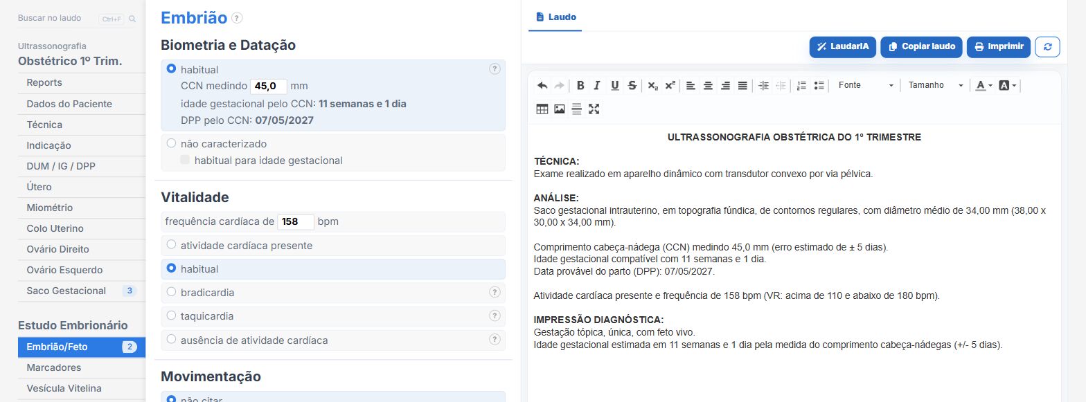
Laudário was developed by and for sonologists precisely to solve these points. For the first-trimester exam, as well as for second- and third-trimester obstetric, morphology, biophysical profile, cervical length, and Doppler, the system offers:
- Ready-made, structured templates for first-trimester obstetric (with and without Doppler), first-trimester morphology, and second- and third-trimester obstetric, organized in tabs (header, indication, gestational sac, yolk sac, embryo, viability, nuchal translucency, morphology, ovaries, corpus luteum, cervix, Doppler, limitations, and conclusion), to speed up typing and prevent omissions.
- Automatic calculations with integrated clinical interpretation: gestational age by CRL and by mean sac diameter, NT percentiles, corrected dating, and estimated due date already integrated into the report, with immediate updating of each block.
- A dedicated nuchal translucency block, integrated with an FMF-type risk calculator, recording NT, fetal heart rate, relevant maternal history, and returning the risk for trisomies 21, 18, and 13 right in the report workflow.
- Standardized, customizable phrases for nasal bone, ductus venosus, tricuspid regurgitation, and other markers, keeping terminology consistent across successive exams of the same patient.
- A pregnancy viability tab that organizes scenarios (viable pregnancy, anembryonic pregnancy, incomplete miscarriage, embryonic demise) with suggested phrases aligned with the objective findings.
- Customized header with the clinic logo and physician details, and a validation QR code on every report, ensuring authenticity and digital traceability.
- A complete library with more than 58 exam templates, covering obstetric, morphology, gynecologic, abdomen, breast, thyroid, small parts, joint, and Doppler exams, accessible online from anywhere and always in sync.
The result is direct: less time typing, fewer forgotten findings, and first-trimester reports that are more consistent across successive exams of the same patient, which makes all the difference in a practice where dating, viability, and early aneuploidy screening must be communicated unambiguously. Instead of starting from a blank page with every patient, you adjust a template that has already been tested, keep the clinic's professional standard, and focus on what really demands your attention: reading the images and integrating with prenatal care.
If you still build your obstetric reports manually or by copying old text, it is worth getting to know Laudário. Try it free for 15 days and see in practice how a well-designed structure saves time on every exam.
The ultrasound reporting platform built by sonologists. We write about reports, classifications and standardization from the real routine of the practice. Who writes and how.
Educational content for healthcare professionals. It does not replace clinical judgment or the guidelines adopted by your service. Editorial policy.