Nuchal Translucency and First-Trimester Screening: How to Measure, Describe Markers and Report Risk
CRL between 45 and 84 mm, NT measurement by FMF criteria, nasal bone, ductus venosus and tricuspid regurgitation, the combined risk and preeclampsia screening, organized in a report the obstetrician reads in seconds.
 LaudárioUltrasound reports in practice
This article is part of the guide Obstetric ultrasound: the three-trimester guide, from dating to Doppler.
The ultrasound between 11 and 14 weeks is no longer just the nuchal translucency exam. Today it is a screening visit: aneuploidy, preeclampsia, definitive dating of the pregnancy and a first anatomic assessment. All of this fits in one report, as long as it is organized so that the obstetrician quickly finds what matters: the measurement, whether it was done correctly and what the resulting risk is.
This text covers how to measure and describe each marker and how to present the risk without turning the report into an unreadable software printout.
The right window and dating
Nuchal translucency (NT) is measured with a crown-rump length (CRL) between 45 and 84 mm, which corresponds roughly to 11 weeks to 13 weeks and 6 days. Outside this range, the measurement cannot be interpreted by the risk models.
The CRL from this exam also defines the dating of the pregnancy. The gestational age established here should be the reference for all subsequent exams, and the report should say so explicitly. It is what will allow weight and Doppler percentiles to be calculated correctly in the third trimester.
How nuchal translucency should be measured
The Fetal Medicine Foundation (FMF) criteria are the standard. In short:
- Midsagittal plane, with the fetus in a neutral position (neither flexed nor hyperextended).
- Magnification such that the head and thorax fill most of the image.
- Distinguish the fetal skin from the amniotic membrane.
- Measure the greatest thickness of the anechoic space, with the calipers placed on the lines (and not inside the space).
- If there is a nuchal cord around the neck, measure above and below and use the average.
The report does not need to describe the technique in detail, but should make clear that the measurement followed the standard and record the number in millimeters with one decimal place. An NT value without the corresponding CRL is of no use.
When the NT is increased
NT increases with gestational age, so interpretation is always by percentile for the CRL. Two practical points:
- Above the 95th percentile for the CRL, the risk of aneuploidy increases and the value enters the risk calculation with significant weight.
- From 3.5 mm (around the 99th percentile), regardless of the calculated risk, there is an indication for workup: offering invasive diagnostic testing or cell-free fetal DNA screening, and fetal echocardiography, because increased NT is also associated with heart defects and genetic syndromes with a normal karyotype.
A report with NT above 3.5 mm should carry this in the diagnostic impression and the recommendation in the recommendations tab, and not just the number.
Additional markers
When assessed by a trained examiner, additional markers refine the risk:
- Nasal bone: present or absent. Absence is associated with trisomy 21, with variation by ethnicity.
- Ductus venosus: positive a wave or absent/reversed. A reversed a wave increases the risk of aneuploidy and of heart disease.
- Tricuspid regurgitation: present or absent.
In the report, each marker is described as assessed or not assessed. "Nasal bone present" and "nasal bone not assessed" are different pieces of information, and the risk calculation changes depending on the presence or absence of the data.
The combined risk
Combined screening brings together maternal age, NT, sonographic markers, fetal heart rate and, when available, free β-hCG and PAPP-A levels. The result is a risk for trisomies 21, 18 and 13, expressed as 1:N.
Good practices for presenting the risk in the report:
- State the baseline risk (by maternal age) and the adjusted risk.
- Say whether the calculation did or did not include biochemistry. A risk without biochemistry has lower performance and this needs to be clear.
- Classify as low, intermediate or high according to the service protocol, and do not just deliver the number.
Preeclampsia screening
The same exam allows screening for early preeclampsia, combining maternal history, mean arterial pressure, the uterine artery PI and, when available, PlGF. High-risk pregnant women benefit from acetylsalicylic acid started before 16 weeks, which gives this screening a direct clinical impact. The report should include the PI of each uterine artery, the mean PI with the multiple of the median and the calculated risk.
Early anatomic assessment
Many major malformations are already visible at this stage: acrania, alobar holoprosencephaly, abdominal wall defects, megacystis, limb abnormalities. A systematic assessment (skull, face, thorax, four-chamber heart when possible, abdomen, bladder, limbs and spine) adds a great deal to the exam, as long as the report makes clear that this is an early assessment and does not replace the second-trimester anatomy scan.
Common errors in the report
- NT without CRL, or CRL outside the 45 to 84 mm range without a caveat.
- Additional markers omitted, without saying whether or not they were assessed.
- Risk without stating whether biochemistry was included.
- NT above 3.5 mm without a recommendation for workup and fetal echocardiography.
- Exam dating not indicated as the reference for the pregnancy.
For the complete structure of the first-trimester exam, see our text on the first-trimester report, from dating to nuchal translucency.
How Laudário helps
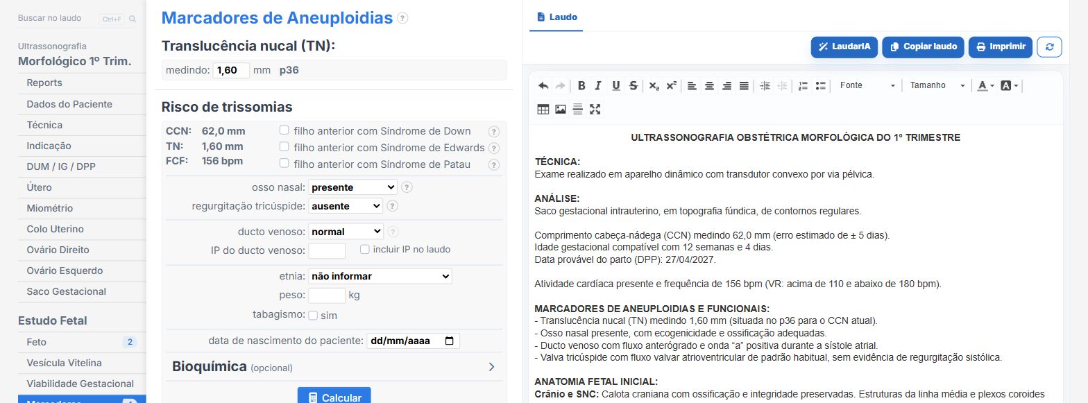
Laudário has first-trimester anatomy scan and obstetric templates, including twin pregnancies. In practice:
- Fields for CRL, NT, nasal bone, ductus venosus and other markers, with a "not assessed" option for each.
- Trisomy and preeclampsia risk calculation integrated into the report, using the Fetal Medicine Foundation algorithms.
- Early anatomic assessment by system.
- Diagnostic impression and recommendations assembled from what was checked.
Try it for 15 days free, no credit card.
The ultrasound reporting platform built by sonologists. We write about reports, classifications and standardization from the real routine of the practice. Who writes and how.
Educational content for healthcare professionals. It does not replace clinical judgment or the guidelines adopted by your service. Editorial policy.