Fetal echocardiography screening: from situs to outflow tracts, with z-scores
The four-chamber view alone detects fewer than half of congenital heart defects. Review the full sequential segmental analysis (situs, four chambers, outflow tracts, three vessels and arches), plus the z-score, the Tei index and the Huhta score.
 LaudárioUltrasound reports in practice
This article is part of the guide Obstetric ultrasound: the three-trimester guide, from dating to Doppler.
Congenital heart disease is the most common malformation at birth (about 8 in every 1,000 live births) and the leading cause of death from congenital anomaly in the first year of life. It is also, historically, the malformation that most often escapes prenatal screening. The reason is well known and has a solution: for decades, cardiac screening was limited to the four-chamber view, and the four-chamber view alone misses precisely the most serious defects: transposition of the great arteries, tetralogy of Fallot, truncus arteriosus. All of them have normal-looking four chambers.
The answer is sequential segmental analysis: a routine that walks through the fetal heart from situs to the outflow tracts and arches, in the same order, in every study. This text walks through that routine, shows what each plane decides, and discusses the tool that turned fetal cardiac biometry from an impression into a measurement: the z-score.
Why four chambers are not enough
The four-chamber view is excellent for what it shows: disproportion between the cavities, atrioventricular septal defects, ventricular hypoplasia, Ebstein anomaly, large effusions. Its stand-alone sensitivity for congenital heart disease, however, is around 40% to 50% in most series.
The reason is anatomic. Conotruncal anomalies (those involving the outflow tract and the relationship between the great vessels) are established above the four-chamber plane. In transposition of the great arteries, the ventricles and atria are normal; what is swapped is where each artery arises. In tetralogy of Fallot, the ventricular septal defect may be subarterial and ventricular disproportion may be minimal until late in pregnancy. In both cases, the four-chamber plane is normal, and a screening that stops there delivers a reassuring report for the fetus that would most need delivery at a tertiary center.
Adding the outflow tracts and the three-vessel view raises detection to the range of 65% to 80% and, in experienced centers, beyond. It is the only protocol change with this impact, and it costs no equipment at all: it costs a routine.
The segmental sequence, plane by plane
The logic of the segmental study is to answer three questions, in this order: where are the viscera and atria (situs)? How do the atria connect to the ventricles (atrioventricular connection)? How do the ventricles connect to the arteries (ventriculoarterial connection)? A heart can have each of these segments normal or abnormal independently, and it is the combination that defines the diagnosis.
Transverse plane of the abdomen: situs
Everything starts below the heart. In the transverse view of the upper abdomen, identify the position of the stomach bubble, descending aorta and inferior vena cava. The four possibilities:
- Situs solitus: stomach bubble and descending aorta on the left, inferior vena cava on the right. The normal pattern.
- Situs inversus totalis: the mirror image. In isolation, it has a low association with heart disease, but it requires checking cardiac position: if the viscera are inverted, the heart should follow. Situs inversus with levocardia is a discordance that heralds complex heart disease.
- Situs ambiguus with right isomerism (asplenia): stomach bubble in a variable position and, the most characteristic sign, inferior vena cava juxtaposed to the descending aorta, on the same side. It is associated with severe heart disease and total anomalous pulmonary venous return.
- Situs ambiguus with left isomerism (polysplenia): the key sign is interruption of the inferior vena cava with azygos continuation. It is associated with complete atrioventricular block.
This is the quickest plane of the study and the one most often skipped. It takes seconds and, when abnormal, reorganizes the interpretation of everything that follows.
Cardiac position and axis
Two different things, often confused. Position describes where the heart is: levocardia, mesocardia, dextrocardia. Dextroposition and levoposition, in turn, describe a heart displaced by an extrinsic cause (diaphragmatic hernia, thoracic mass, pleural effusion) and not an anomaly of laterality. Distinguishing the two is essential: dextrocardia is a cardiac problem; dextroposition is a normal heart pushed by a left diaphragmatic hernia.
The cardiac axis has a reference of approximately 45° ± 20° (range 25° to 65°). It is an underrated marker: axis deviation, especially to the left, is associated with conotruncal anomalies and diaphragmatic hernia, and is often the first abnormal thing in a heart that looks normal.
The cardiac area is assessed by the ratio of cardiac to thoracic circumference (CC/TC), with a reference of less than 0.55: the normal heart occupies less than one third of the thoracic area. Values of 0.55 or more indicate cardiomegaly and warrant investigation of the cause: volume overload, cardiomyopathy, fetal anemia, arteriovenous fistula.
Four-chamber plane
Here the analysis is systematic. What should be included:
- General appearance, with an important reminder: right-sided cavity predominance is expected in the fetal circulation. A right ventricle slightly larger than the left is normal, and calling this disproportion generates unnecessary referrals. What is concerning is significant asymmetry, or left predominance (which is abnormal at any gestational age).
- Individual chambers: atria and ventricles, each normal, enlarged or hypoplastic; for the ventricles, also attention to wall hypertrophy.
- Atrial septum and foramen ovale: normally functioning, restrictive, prematurely closed, or with an atrial septal defect. It is worth noting that the diagnosis of ASD in the fetus is intrinsically limited, since the foramen ovale is a physiologic communication, and the report should include this caveat.
- Interventricular septum: intact within the resolution limits of the method, or with a VSD, and here location matters a great deal: perimembranous, muscular, subpulmonary (subarterial), inlet or multiple. A subarterial VSD points to the conotruncal spectrum; an inlet VSD points to atrioventricular septal defect.
- Atrioventricular valves: morphology and function on color flow mapping, with attention to the normal offset between the tricuspid and mitral valves (the tricuspid inserts more apically).
- Pericardial effusion: graded in millimeters: minimal/physiologic (less than 2 mm), small (2 to 4 mm), moderate (5 to 9 mm) or large (10 mm or more, with potential for hemodynamic compromise).
One finding in this plane deserves special note: absence of the cardiac crux, with a common atrioventricular valve, ostium primum ASD and inlet VSD. This is the complete atrioventricular septal defect, and it should be described as a single, cohesive entity, not as four isolated findings, because that is what it is. Describing “ASD”, “VSD”, “tricuspid abnormality” and “mitral abnormality” on separate lines fragments a diagnosis that has its own name and a strong association with trisomy 21.
Outflow tracts: the plane that changes the outcome
The question is a single one: does the aorta arise from the left ventricle and the pulmonary trunk from the right ventricle, crossing each other just after their origin? The crossing is the point. Two arteries that emerge in parallel, without crossing, are a transposition until proven otherwise.
The ventriculoarterial connection possibilities that the report should be able to name:
- Concordant: the normal pattern, with preserved mitral-aortic continuity.
- Transposition of the great arteries (TGA): aorta from the right ventricle, parallel to the pulmonary trunk.
- Aortic override: tetralogy of Fallot: the aorta arises from the left ventricle overriding the interventricular septum.
- Double outlet right ventricle (DORV) or double outlet left ventricle.
- Truncus arteriosus: a single artery emerges from the heart; the aorta cannot be identified separately.
- Pulmonary atresia, with intact septum or with VSD (tetralogy spectrum).
- Aortic atresia.
- Corrected transposition (CCTGA): the double discordance, atrioventricular and ventriculoarterial.
A rigor worth adopting: do not state a normal dimension without having measured it. Writing “ascending aorta of normal caliber” without a measurement is an unsupported statement, and it is exactly where mild hypoplasia escapes.
Three vessels and three vessels-trachea (3V and 3VT)
Probably the plane with the best effort-to-yield ratio of the entire screening. The normal rule is a relationship of calibers: PA > Ao > SVC, arranged in a line, with the trachea to the right of the aortic arch.
What this plane delivers:
- Pulmonary artery of reduced caliber relative to the aorta: tetralogy of Fallot, pulmonary atresia, pulmonary stenosis.
- Aorta of reduced caliber relative to the pulmonary artery: coarctation, hypoplastic left heart, aortic atresia.
- Vessels in parallel: transposition.
- Aneurysmal pulmonary trunk and branches: absent pulmonary valve syndrome.
- Right aortic arch, double arch, aberrant right subclavian artery: arch anomalies, with tracheal deviation as an indirect sign. The double arch constitutes a vascular ring and has postnatal respiratory implications.
- Ductal and aortic convergence into the descending aorta in the “V” pattern, and color inversion between the two vessels, which reveals reverse flow.
The aortic isthmus deserves specific attention because of coarctation, the heart defect with the highest prenatal false-negative rate: the patent ductus arteriosus masks the obstruction until birth. An isthmus of reduced caliber raises the risk and justifies serial follow-up, even though the definitive diagnosis often comes only after ductal closure.
Z-score: when an impression becomes a measurement
“Left ventricle of reduced dimensions for gestational age” is a phrase that means different things at 22 and at 34 weeks. The fetal heart grows, and every judgment of caliber needs to be relative to gestational age. That is what the z-score does: it expresses how many standard deviations the measurement is from the mean expected for that gestational age.
The most widely used model is log-normal: Z = (ln(x) − ln(mean)) / sigma, with means and sigmas tabulated week by week. The established references are Schneider et al. (2005) for cardiac structures and Pasquini et al. (2007) for the aortic isthmus and ductus arteriosus.
The structures that benefit from the z-score, grouped by segment:
- Left heart: mitral annulus, LV width and length, aortic annulus, ascending aorta, descending aorta.
- Right heart: tricuspid annulus, RV width and length, pulmonary annulus, pulmonary trunk, right and left pulmonary artery branches.
- Isthmus and ductus: aortic isthmus in the three-vessel view, ductus arteriosus in the three-vessel view, aortic isthmus in the sagittal view.
The interpretation is straightforward:
- |Z| ≤ 2: normal.
- 2 < |Z| ≤ 3: borderline.
- |Z| > 3: abnormal.
Two cautions. The first: the z-score is descriptive, not diagnostic. It tells you that a dimension is outside the range; it does not tell you why. A mature report states “pulmonary valve with markedly reduced dimension (Z −5.80)” and leaves the diagnosis to clinical synthesis, rather than deducing the heart defect from the number.
The second: the z-score depends entirely on gestational age. A wrong GA produces wrong z-scores with an appearance of precision. Before trusting a z-score, check the dating, especially in pregnancies without an early ultrasound.
One additional practical caution: very extreme values (|Z| above 4, and especially above 8) deserve rechecking of the measurement before going into the report. Not because they are impossible, but because a typing or caliper error produces exactly this pattern.
Rhythm, velocities and function
Rhythm
The normal fetal heart rate is between 120 and 180 bpm, with 1:1 rhythm (each atrial contraction followed by a ventricular one). The report should record the ventricular rate and, when the rhythm is not 1:1, the atrial rate as well, because the relationship between the two is the diagnosis.
- Extrasystoles: the most common fetal arrhythmia and almost always benign. It is worth characterizing origin (atrial, ventricular), conduction (conducted, non-conducted/blocked, mixed) and frequency (isolated, frequent, bigeminy, trigeminy). Frequent blocked atrial extrasystoles can mimic bradycardia.
- Tachyarrhythmias: supraventricular tachycardia (typically 1:1, around 220 to 260 bpm), atrial flutter (atrial rate of 300 to 500 bpm with variable conduction), atrial fibrillation and ventricular tachycardia. The distinction defines transplacental maternal treatment and is urgent, because sustained tachyarrhythmia leads to hydrops.
- Bradyarrhythmias and atrioventricular block: in complete AV block, the hallmark is complete atrioventricular dissociation, with normal atrial rate and slow ventricular rate. It has two associations that must be actively investigated: maternal anti-Ro/anti-La antibodies and structural heart disease, particularly left isomerism.
The atrioventricular conduction time, measured in the 3VT view, has an approximate reference of 100 to 140 ms; above 150 ms suggests first-degree block. It is the measurement that allows AV block to be detected in its early phase, precisely in the at-risk population (pregnant patients with anti-Ro), when intervention can still change the course.
Doppler velocities
Reference values that guide the reading of the outflow tracts and the ductus:
- Aortic valve: 60 to 120 cm/s; above 200 cm/s, aortic stenosis.
- Pulmonary valve: 70 to 130 cm/s; above 200 cm/s, pulmonary stenosis.
- Ductus arteriosus, systolic: 50 to 180 cm/s. The ductus physiologically has the highest velocity in the fetal heart.
- Ductus arteriosus, diastolic: 8 to 30 cm/s.
The gradient is derived by the simplified Bernoulli equation (ΔP = 4V²).
Two readings deserve emphasis. Ductal constriction (systolic above 180 cm/s or diastolic above 30 cm/s) is often iatrogenic, caused by nonsteroidal anti-inflammatory drugs taken by the mother, and is reversible once the drug is stopped. It is one of the few findings in which the report can literally solve the problem by naming it.
Tricuspid regurgitation has a significance criterion that combines two parameters: velocity above 200 cm/s and duration longer than 70 ms. Low-velocity, short-duration jets are frequent and nonspecific findings. The mitral jet, on the other hand, has a counterintuitive reading: a high jet (300 to 400 cm/s) is physiologic, because it reflects the normal gradient between LV and LA; the finding is the very existence of regurgitation. A low mitral jet, in contrast, suggests left ventricular dysfunction.
Function
Two tools deserve to be in the report when the fetus has any sign of compromise:
The Tei index (myocardial performance index) combines the isovolumetric contraction and relaxation times and the ejection time: Tei = (IVRT + IVCT) / ET. Approximate references: LV ≤ 0.50 and RV ≤ 0.55. It is a global index, sensitive early and independent of ventricular geometry, which is useful precisely in malformed hearts, where geometric measures of function fail.
The Huhta cardiovascular score organizes hemodynamic repercussion into 5 domains of 2 points each, totaling 10:
- Hydrops: absent (2); ascites or effusion (1); overt hydrops (0).
- Venous Doppler: ductus venosus with positive A wave and umbilical vein without pulsations (2); absent A wave or pulsatile umbilical vein (1); reversed A wave with pulsatile umbilical vein (0).
- Heart size: CC/TC less than 0.35 (2); 0.35 to 0.50 (1); above 0.50 or below 0.20 (0).
- Cardiac function: normal biventricular without regurgitation (2); mild dysfunction or holosystolic regurgitation (1); severe dysfunction or severe regurgitation (0).
- Umbilical artery: normal diastolic flow (2); absent diastolic flow (1); reversed diastolic flow (0).
The interpretation: 10 is normal; 7 or less indicates mild compromise; 5 or less, moderate to significant compromise; below 5, severe compromise, with high mortality. It is a score that translates the study into urgency, and therefore directly routes the referral.
Indications: who should have fetal echocardiography
Basic cardiac screening belongs to the anatomy scan of every pregnant patient. Dedicated fetal echocardiography has its own indications:
- Suspected structural heart disease on the anatomy scan: the most frequent indication.
- Congenital heart disease in a previous child or a first-degree relative.
- Pregestational diabetes mellitus.
- Maternal systemic lupus erythematosus, or anti-Ro/anti-La antibodies.
- Use of teratogenic drugs or medications.
- First-trimester viral infection.
- Increased nuchal translucency (≥ 3.5 mm), even with a normal karyotype: one of the most underused indications.
- Fetal chromosomal abnormality.
- Fetal hydrops.
- Fetal arrhythmia identified prenatally.
- Monochorionic twin pregnancy.
- Intrauterine growth restriction.
Pitfalls in the fetal echocardiography report
- Concluding normality from the four chambers: the origin of most serious false negatives.
- Calling physiologic right predominance a disproportion: generates unnecessary referrals.
- Stating normal caliber without a measurement: this is where mild hypoplasia escapes.
- Trusting a z-score with doubtful GA: the precision is apparent; the error is real.
- Reading the z-score as a diagnosis: it reports dimension, not disease.
- Fragmenting AVSD into four isolated findings instead of naming the entity.
- Not distinguishing dextrocardia from dextroposition: they are completely different problems.
- Omitting the caveat about ASD, VSD and coarctation: these are the three recognized limitations of the method in the fetal period, and the report should say so.
- Not recording the atrial rate in arrhythmias: the atrioventricular relationship is the diagnosis.
- Failing to name ductal constriction: a reversible finding whose cause is usually in the prescription.
How Laudário helps structure fetal echocardiography
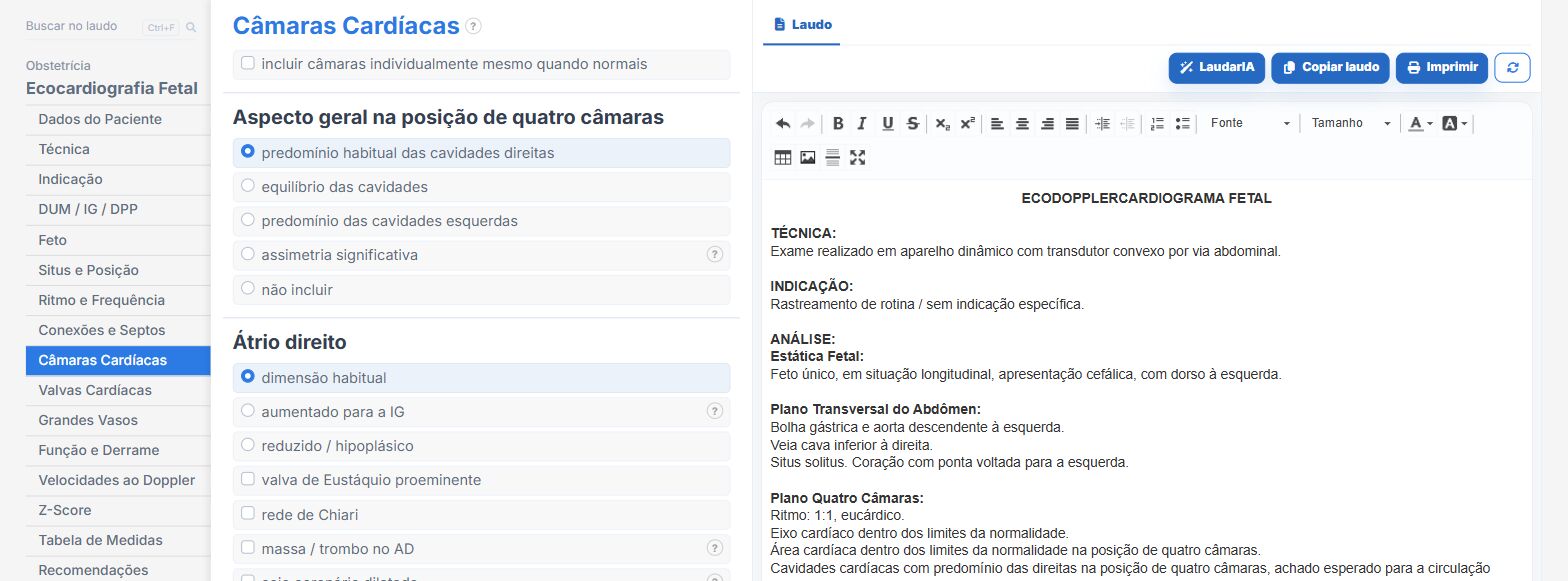
Laudário has a complete fetal echo-Doppler cardiography module, built on the segmental sequence. In practice, this means:
- Report assembled in scanning order (abdomen and situs, four chambers, LV outflow, RV outflow, three vessels and 3VT, aortic arch, ductal arch, function), so that the form’s routine is the study’s routine.
- Automatic consistency between segments: the text for the stomach bubble, vena cava and descending aorta is derived from the chosen situs, and the system flags the discordance between situs inversus and cardiac position.
- Z-score engine with 16 cardiac structures, based on Schneider (2005) and Pasquini (2007), with interpolation by gestational age, the p5-p95 reference range displayed beside the field, automatic coloring for normal, borderline and abnormal ranges, and an alert for physiologically implausible values.
- Automatic impression for structures with |Z| > 2, written in a descriptive and graded way (mildly, no adverb, markedly), without jumping to the diagnosis.
- Automatic calculation of the CC/TC ratio with a cardiomegaly alert, the Tei index from the times, the gradient by Bernoulli and the Huhta score, with the score automatically routing the urgency of the referral.
- Cohesive descriptions for entities that should not be fragmented: AVSD comes out as a single paragraph, with automatic suppression of the isolated ASD, VSD and valve lines, because they are components of the canal itself.
- Thirty ready-made heart disease templates (HLHS, tetralogy of Fallot, TGA, CCTGA, AVSD, coarctation, Ebstein, truncus arteriosus, fetal SVT, complete AV block, rhabdomyoma, vascular ring, NSAID-induced ductal constriction, among others), which fill in a coherent segmental constellation with one click, and which should, naturally, be reviewed before finalizing.
- A velocities tab that mirrors the valve fields, allowing block entry without losing any of the automatic classifications.
- Fixed footer caveat about the recognized limitations of the method (ASD, VSD and coarctation, due to the peculiarities of the fetal circulation) and automatically printed references (Donofrio et al., Circulation 2014; Schneider et al., 2005).
- Support from LaudarIA, the integrated artificial intelligence, to assist in writing complex heart defects and cases with multiple affected segments.
The result is a report that gives the pediatric cardiologist what they need: anatomy segment by segment, measurements with z-score, quantified hemodynamic repercussion and explicit urgency of referral. In a condition where prenatal diagnosis changes neonatal mortality (because it defines the place of delivery and the availability of cardiac surgery in the first hours of life), the structure of the report has a direct consequence on the outcome.
If you perform fetal echocardiography and still build the report manually, it is worth trying: Laudário offers a 15-day free trial, no credit card required, with immediate access to the fetal echo-Doppler cardiography module, the first-, second- and third-trimester anatomy and growth studies and all the other protocols of the obstetric routine.
The ultrasound reporting platform built by sonologists. We write about reports, classifications and standardization from the real routine of the practice. Who writes and how.
Educational content for healthcare professionals. It does not replace clinical judgment or the guidelines adopted by your service. Editorial policy.