Cervical length measurement in obstetric ultrasound: how to measure, stratify risk and standardize the report
How to perform and structure the transvaginal cervical length report in the second trimester: standardized technique, cutoffs for preterm birth risk, assessment of the internal os and funneling, and how to keep serial exams consistent.
 LaudárioUltrasound reports in practice
This article is part of the guide Obstetric ultrasound: the three-trimester guide, from dating to Doppler.
Cervical length measurement has gone from being a complementary exam to one of the most important tools for screening and preventing preterm birth. In a country where preterm birth is still among the leading causes of neonatal mortality, measuring the cervix reproducibly and structuring a clear report is a direct clinical responsibility of the sonologist. The exam is simple, fast and inexpensive, but it only delivers real value when it follows a protocol, provides reliable measurements and ends with an objective conclusion about risk.
In practice, however, there is considerable heterogeneity among services: reports with isolated measurements and no gestational context, incomplete descriptions of the internal os, no information about funneling, and generic conclusions such as “cervix within normal limits”. This compromises obstetric decision-making and, ultimately, the quality of care given to the pregnant woman. In this article, we lay out a practical structure for cervical length assessment, from technique to the final report, and show how to standardize this exam in your routine.
Why cervical length matters
Cervical length between 18 and 24 weeks is the best single predictor of spontaneous preterm birth in singleton pregnancies. The shorter the cervix, the higher the risk, and this relationship is continuous, with no single cutoff. Even so, the literature and the main consensus statements (FMF, SMFM, FEBRASGO) converge on some practical references: a cervix equal to or shorter than 25 mm in a singleton pregnancy in the second trimester identifies a high-risk pregnant woman and justifies preventive measures, such as vaginal progesterone or cerclage in selected cases.
There is therefore a direct clinical consequence to every report issued. An inaccurate measurement may lead to an unnecessary intervention or, worse, fail to identify a pregnant woman who would benefit from treatment. Standardizing the method is what makes cervical length reproducible across professionals, services and over time in the same patient.
When to order cervical length measurement
There are two main indication scenarios. The first is universal second-trimester screening, performed between 20 and 24 weeks, usually combined with the anatomy scan, with the goal of identifying asymptomatic pregnant women with a short cervix. The second is serial surveillance in high-risk patients (history of spontaneous preterm birth, multiple gestations, uterine malformations, prior conization, confirmed cervical insufficiency), in whom the exam is repeated every one to two weeks, typically between 16 and 24 weeks.
Outside these contexts, cervical length is also requested in specific situations, such as evaluation of threatened preterm labor, post-cerclage monitoring and investigation of second-trimester bleeding. Each scenario changes the focus of the report: screening looks for the risk cutoff; surveillance looks for the shortening curve over time; symptomatic evaluation looks for dynamic findings.
Standardized technique: what cannot be missing
The transvaginal route is the gold standard. Although the transabdominal and transperineal routes can be used in specific situations, both tend to overestimate cervical length and have lower reproducibility. Before the measurement, the patient should empty her bladder: a full bladder compresses the cervix and can mask funneling, producing a falsely normal measurement by up to 5 mm.
The acquisition technique follows a few essential rules that need to be built into the routine:
- Probe placed in the anterior fornix, without excessive pressure on the cervix.
- Simultaneous visualization of the internal os, the external os and the endocervical canal in a sagittal view.
- Image occupying at least 75% of the screen.
- Anterior and posterior cervical lips of similar thickness (avoid oblique images).
- Three measurements acquired; record the shortest.
- Observation for at least three to five minutes to detect dynamic funneling.
- Provocative maneuvers (gentle fundal pressure, Trendelenburg position) in selected cases.
The measurement should be linear, between the internal and external os, following the endocervical canal. When the cervix is curved, two straight lines can be added together, avoiding the underestimation that occurs when a single straight line is drawn between the os. This technical detail is often forgotten and is a major source of interobserver variation.
Cutoffs and risk stratification
Interpretation of cervical length must be tied to gestational age and the patient's risk profile. In a singleton pregnancy in the second trimester, the most commonly used references are:
- Cervix > 25 mm: within the normal range.
- Cervix between 20 and 25 mm: increased risk; consider vaginal progesterone and follow-up.
- Cervix < 20 mm: significantly elevated risk; clear indication for vaginal progesterone; evaluate cerclage in selected patients.
- Cervix < 15 mm: high risk of spontaneous preterm birth, even in patients with no obstetric history.
In twin pregnancies, the cutoff is usually shifted to 25 mm as a threshold for attention, and measurements below 20 mm warrant multidisciplinary discussion. In patients with a history of preterm birth, any progressive shortening between serial exams is relevant, even if the absolute value is not below the cutoff.
Besides the measurement, the report needs to characterize funneling of the internal os: its presence, shape (T, Y, V, U) and depth. Isolated funneling, without shortening of the functional cervix, has limited predictive value, but it reinforces the risk when associated with a short cervix.
Structure of a complete report
A standardized cervical length report should include, at minimum, seven blocks of information. The absence of any of them makes comparison with serial exams difficult and weakens the conclusion.
1. Identification and gestational age. Gestational age by last menstrual period and by biometry; corrected gestational age when there is a significant discrepancy. Interpretation of the cutoffs changes with gestational age.
2. Route of examination and technical conditions. Record the transvaginal route, bladder status, patient position and any limitations (scar, discomfort, anatomical changes).
3. Description of the cervix. Appearance of the endocervical canal, echogenicity, presence of Nabothian cysts, integrity of the internal and external os.
4. Functional length measurement. Value in millimeters, referencing the shortest of three measurements. Record the resting measurement and the measurement after a provocative maneuver separately, if performed.
5. Assessment of the internal os and funneling. Presence, shape and depth. When absent, record “internal os closed, no funneling”.
6. Dynamic assessment. Observation time and response to provocative maneuvers, when applicable.
7. Objective conclusion. Classification of the cervix (normal, short, very short) with reference to the cutoff used, considering gestational age and clinical context. Recommend reassessment when applicable.
Common pitfalls that compromise the report
Although technically simple, a few recurring errors undermine the quality of cervical length measurement in routine practice. A full bladder is the most common: it overestimates length and masks funneling, producing false negatives. Excessive probe pressure on the cervix is another classic problem: it can stretch the endocervical canal and artificially increase the measurement by 4 to 5 mm. A single measurement without dynamic assessment is a trap that ignores the physiology of the cervix, which can shorten within a few minutes under stimulation.
Other points of attention include acquisition in an oblique plane (which apparently shortens the length), failure to recognize the true internal os in cervices with an already distended lower segment, and failure to describe funneling when it is present. Finally, generic conclusions such as “within normal limits for gestational age” that omit the exact value and the cutoff used impair follow-up and, in borderline cases, may lead to inappropriate clinical decisions.
How Laudário helps standardize cervical length reporting
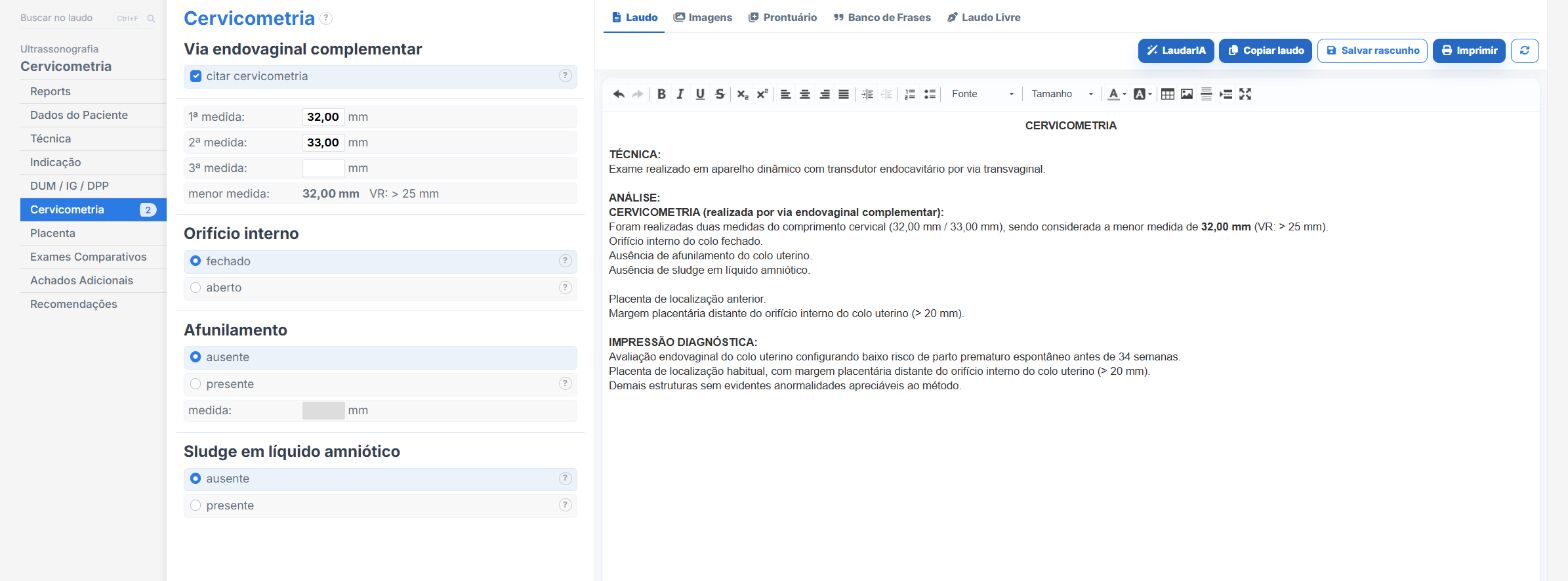
The real challenge of cervical length assessment is not the technique itself: it is keeping, exam after exam and patient after patient, the same report structure, with every item filled in and an objective conclusion. This is exactly where Laudário fits into the routine. The system offers a dedicated cervical length template, with a ready-made structure to describe the route of examination, technical conditions, functional length, funneling and dynamic assessment, so the sonologist does not have to rewrite the skeleton of the report for every exam.
With customizable phrases, pre-built conclusions and a workflow designed for the reality of obstetric ultrasound, Laudário drastically reduces typing time and eliminates the most common oversights, such as forgetting to cite the cutoff, omitting the dynamic assessment or forgetting to classify the internal os. In patients under follow-up, this makes it easier to compare serial exams, because all reports follow the same structure and the same language.
If your routine involves cervical length measurement, whether as a standalone exam or combined with the second-trimester anatomy scan, it is worth seeing Laudário in practice. The system offers a free 15-day trial, no credit card required, with immediate access to the entire library of ultrasound templates. In just a few exams you will notice the difference a standardized report makes in productivity, clinical consistency and the quality delivered to the patient.
The ultrasound reporting platform built by sonologists. We write about reports, classifications and standardization from the real routine of the practice. Who writes and how.
Educational content for healthcare professionals. It does not replace clinical judgment or the guidelines adopted by your service. Editorial policy.