Baby's sex on ultrasound: how many weeks it is reliable and how to record it in the report
The genital tubercle angle in the first trimester, direct visualization from 16 weeks, the most common errors, respecting those who do not want to know, and atypical genitalia, which stops being a curiosity.
 LaudárioUltrasound reports in practice
This article is part of the guide Obstetric ultrasound: the three-trimester guide, from dating to Doppler.
"How many weeks can you see the baby's sex on ultrasound?" is one of the questions patients ask Google most, and one the sonologist answers most often in the exam room. For the physician, the question is different: from what point is the information reliable enough to go into the report, and how to write it so as not to cause disappointment at the gender reveal party.
This text summarizes what the literature shows about accuracy at each stage, how to describe the genitalia and when the subject stops being a curiosity and becomes an investigation.
First trimester: the genital tubercle angle
Between 11 and 14 weeks, the external genitalia are not yet differentiated. The method used is the genital tubercle angle in the midsagittal view:
- An angle above 30 degrees relative to a horizontal line through the lumbosacral spine suggests male sex.
- A parallel or converging tubercle (below 10 degrees) suggests female sex.
- Between 10 and 30 degrees, indeterminate.
Accuracy improves with gestational age: it is low at 11 weeks and reaches high values near 13 weeks, but depends heavily on the view and the examiner, and errors are more frequent for male sex. For this reason, in this period the information should be given as probable, and the report should make this clear, if the practice chooses to record it.
Second trimester: direct visualization
From about 16 weeks, the external genitalia can be seen directly, and accuracy approaches 100% under good technical conditions. In the second-trimester anatomy scan, between 20 and 24 weeks, the assessment is very reliable.
- Male: penis and scrotum visible in axial and sagittal views.
- Female: labia majora and minora, in the axial view (the "three lines" sign).
Errors in this phase usually come from an unfavorable fetal position, the umbilical cord between the legs (mistaken for a penis) or swollen labia (mistaken for a scrotum). When it is not possible to see safely, the report should say "genitalia not characterized due to fetal position", and not take a risk.
The laboratory alternative
Sex determination by cell-free fetal DNA in maternal blood, from 8 to 10 weeks, has high accuracy and is currently the earliest way to know the sex. It is worth mentioning to the patient when she is in a great hurry, because it avoids pressure on a first-trimester study whose purpose is something else.
How to record it in the report
- First trimester: if recorded, as "probable fetal sex", with the caveat of limited accuracy at this stage.
- Second trimester: "external genitalia of male appearance" or "female", after direct visualization.
- When it was not possible: "genitalia not characterized", with the reason.
- Respect the wishes of the pregnant patient who does not want to know: many practices record only "external genitalia of usual appearance" in the report, so that the sex is not revealed by whoever reads the document.
This last point is often forgotten. A report can be read by family members before the pregnant patient decides to share the news. Asking and recording the preference avoids problems.
When it stops being a curiosity: atypical genitalia
Ultrasound can also show ambiguous or atypical genitalia, and then the description needs to be careful:
- Hypospadias: the "tulip" sign (short, curved penis between the scrotal folds).
- Clitoromegaly and labial fusion.
- Micropenis.
- Discordance between the sex by fetal DNA or karyotype and the appearance of the genitalia on ultrasound.
In these cases, the report should describe what was seen, without assigning a sex, and recommend evaluation in maternal-fetal medicine and genetic counseling. Congenital adrenal hyperplasia and chromosomal abnormalities are among the causes.
Common errors
- Stating the sex in the first trimester without the probability caveat.
- Mistaking the cord for a penis and swollen labia for a scrotum.
- Taking a risk when the genitalia were not well seen.
- Writing the sex in the report of a patient who asked not to know.
- Assigning a sex to atypical genitalia.
See also our article on the second-trimester anatomy scan.
How Laudário helps
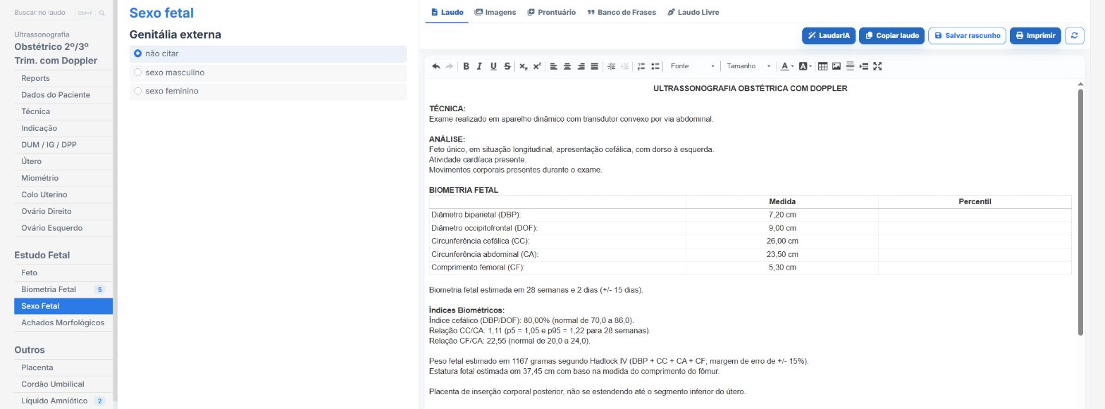
In the obstetric and anatomy scan templates of Laudário:
- External genitalia of male or female appearance, with its own phrase.
- Genitalia with atypical development described without assigning a sex, when characterization is not possible.
- Recommendations in their own tab.
Try it free for 15 days, no credit card required.
The ultrasound reporting platform built by sonologists. We write about reports, classifications and standardization from the real routine of the practice. Who writes and how.
Educational content for healthcare professionals. It does not replace clinical judgment or the guidelines adopted by your service. Editorial policy.