Pancreas ultrasound: acute and chronic pancreatitis, cysts and what to write in the report
The pancreas is the organ that ultrasound sees only halfway most often. How to describe the limitations, look for the biliary cause of pancreatitis, classify collections by the revised Atlanta classification, and what to recommend for a cyst.
 LaudárioUltrasound reports in practice
This article is part of the guide Abdominal Ultrasound: A Guide to the Findings That Actually Change Management.
The pancreas is the upper abdominal organ that ultrasound most often sees only halfway. Retroperitoneal, behind the stomach and gas-filled bowel loops, it usually appears well in the head and body and poorly in the tail. Even so, the exam has a real role: in acute pancreatitis, it looks for the biliary cause and complications; in chronic pancreatitis, it shows calcifications and ductal dilatation; and many pancreatic cysts are incidental findings on routine ultrasound.
This text organizes what to describe in each scenario and, above all, how to write the limitations so that a partially seen pancreas is not read as a normal pancreas.
Reference anatomy and technique
Vascular landmarks locate the organ: the splenic vein runs posterior to the body and tail; the splenomesenteric confluence lies behind the neck; the superior mesenteric artery and the inferior vena cava help identify the head and the uncinate process.
Maneuvers that improve visualization:
- Adequate fasting, to reduce gas.
- Deep inspiration, which lowers the liver and uses it as an acoustic window.
- Lateral decubitus and sitting positions.
- Water ingestion, which distends the stomach and turns gas into a fluid window.
The report should state which segments were evaluated: "head and body visualized; tail partially characterized due to bowel gas interposition". This sentence prevents the referring physician from concluding that the tail is normal.
The normal pancreas and fatty replacement
Pancreatic echogenicity is equal to or greater than that of the liver and increases with age. Fatty replacement (diffusely hyperechoic pancreas, with normal or reduced volume) is a common finding in the elderly, the obese and diabetics, and in most cases has no clinical significance. It is worth describing it as such, without turning it into disease.
The normal main pancreatic duct (Wirsung) measures up to about 3 mm in the body. When visible, the measurement should be in the report.
Acute pancreatitis
The diagnosis is clinical and laboratory-based; ultrasound is not needed to confirm it. The role of the exam is different:
- Look for the biliary cause: cholelithiasis, microlithiasis, biliary sludge and common bile duct dilatation. This is the most important question of the exam, because it determines cholecystectomy and, sometimes, endoscopic cholangiography.
- Describe the pancreas: enlargement, diffuse hypoechogenicity, blurred contours. Early on, the pancreas can be normal on ultrasound, and this does not exclude pancreatitis.
- Identify complications: peripancreatic fluid and collections.
For collections, the revised Atlanta classification separates the situations according to time and content:
- Acute peripancreatic fluid collection: in the first 4 weeks, without a defined wall.
- Pseudocyst: after 4 weeks, a collection with a well-defined wall and fluid content.
- Acute necrotic collection and walled-off necrosis: when there is necrosis, with heterogeneous content and debris.
Ultrasound is limited in distinguishing necrosis from fluid; contrast-enhanced CT is the reference exam for this. The report should describe the size, location and content of the collections and, if there is doubt about necrosis, recommend CT.
Chronic pancreatitis
- Parenchymal calcifications and intraductal stones, hyperechoic with shadowing.
- Irregular dilatation of the Wirsung duct, in a "chain of lakes" pattern.
- Atrophy and irregular contours.
- Associated pseudocysts.
Wirsung duct dilatation deserves care: it may be chronic pancreatitis, but it may also be obstruction by a neoplasm of the head. Simultaneous dilatation of the pancreatic duct and the common bile duct (the "double duct" sign) is a red flag for a periampullary lesion and must be in the diagnostic impression.
Solid focal lesions
Adenocarcinoma usually appears as a hypoechoic, poorly defined mass, often in the head, with upstream ductal dilatation. Small lesions can be isoechoic and go unnoticed, visible only through ductal dilatation or an altered contour. Neuroendocrine tumors tend to be better delimited and more vascular.
When faced with a solid pancreatic mass, the report describes location, measurements, relationship to the vessels (mesenteric, celiac trunk, portal vein), ductal dilatation and secondary signs (lymph nodes, liver lesions, ascites) and recommends staging with CT or MRI.
Pancreatic cysts
With the increasing use of imaging, incidental pancreatic cysts are frequent, especially in the elderly. Ultrasound rarely characterizes the cyst type on its own, but the report can describe the elements that guide the next step:
- Size, location and number.
- Walls, septa, solid component or mural nodule.
- Communication with the main duct, when visible.
- Wirsung duct caliber: dilatation of the main duct, especially from 5 mm, is one of the worrisome features in the guidelines for intraductal papillary mucinous neoplasm (IPMN).
The usual recommendation for an incidental pancreatic cyst is characterization by magnetic resonance with cholangiopancreatography, and the report should carry this recommendation in its own tab.
Common errors
- Writing "normal pancreas" when only the head was seen.
- Acute pancreatitis without detailed evaluation of the gallbladder and common bile duct.
- Collection without measurements and without temporal classification.
- Dilated Wirsung duct without considering obstruction.
- Pancreatic cyst without a recommendation for characterization.
For the context of abdominal findings that change management, see our abdominal ultrasound guide.
How Laudário helps
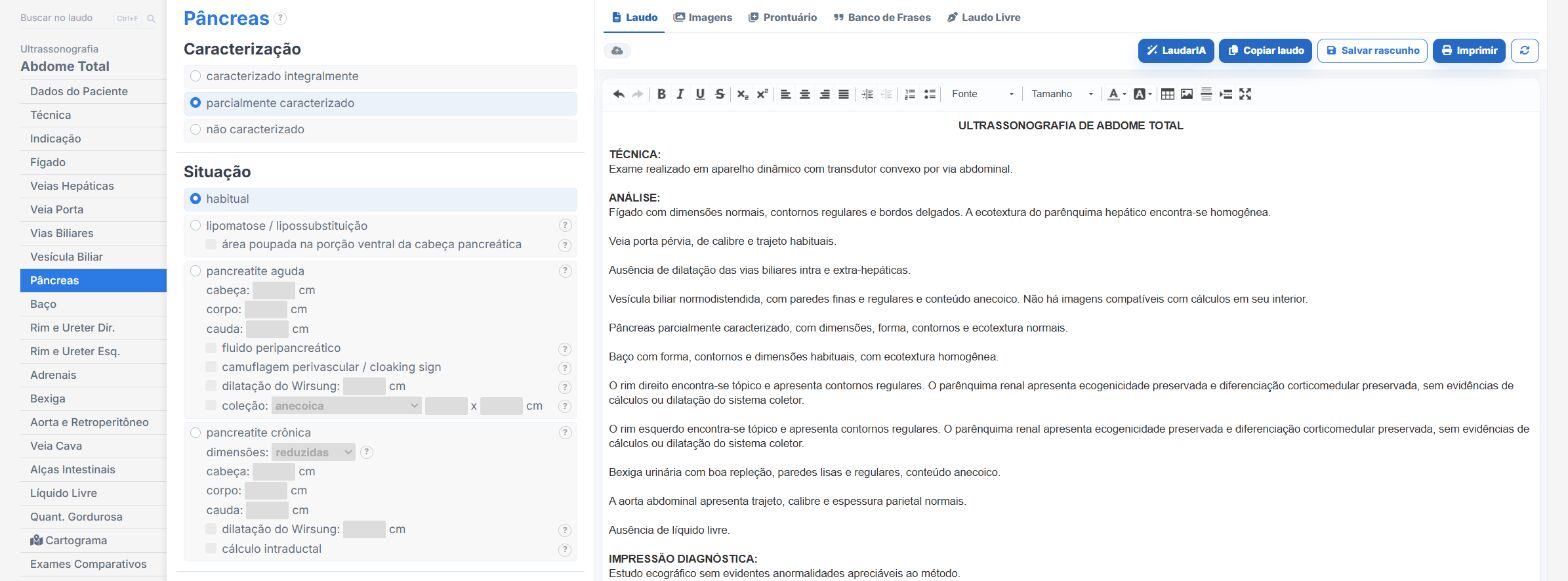
In the abdomen templates of Laudário, the pancreas has its own tab. In practice:
- Option for bowel gas interposition and partial visualization, which enters the report as a limitation.
- Fatty replacement, pancreatitis, pseudocyst and Wirsung duct dilatation with ready-made phrases.
- Gallbladder and bile ducts in the same report, in their own tabs, so the biliary cause is never left out.
- Recommendations in a separate tab.
Try it for 15 days free, no credit card required.
The ultrasound reporting platform built by sonologists. We write about reports, classifications and standardization from the real routine of the practice. Who writes and how.
Educational content for healthcare professionals. It does not replace clinical judgment or the guidelines adopted by your service. Editorial policy.