Spleen and abdominal lymph node ultrasound: splenomegaly, focal lesions and what to measure
How to measure the spleen and relate splenomegaly to the rest of the exam, tell an accessory spleen from a nodule, describe infarct, abscess and lymphoma, and measure lymph nodes by the short axis.
 LaudárioUltrasound reports in practice
This article is part of the guide Abdominal Ultrasound: A Guide to the Findings That Actually Change Management.
The spleen usually takes up one line of the abdominal report: "spleen of normal size and echotexture". Most of the time that is right. But when it is enlarged, when a nodule appears or when there are lymph nodes in the abdomen, the report needs measurements, criteria and context, because these findings point to systemic diseases such as portal hypertension, hematologic diseases, infections and lymphoma.
This text organizes the evaluation of the spleen and abdominal lymph nodes and shows how to write each finding in a useful way.
How to measure the spleen
The most commonly used measurement is the longitudinal length (longest axis), obtained in a coronal view through the left intercostal window, with the patient in the right lateral decubitus position. Practical references in adults:
- Up to about 12 cm: normal.
- Between 12 and 13 cm: borderline, to be interpreted according to body type and height (tall men may have a larger spleen without disease).
- Above 13 cm: splenomegaly.
Some practices also use the splenic index (product of the three diameters) or the estimated volume, which are more reproducible in follow-up. Whatever the method, the report should give the number, and not just "enlarged spleen". In children, the reference is the age-based table.
Splenomegaly: the clue is in the rest of the exam
Splenomegaly is rarely an isolated finding. The report gains value when it relates the enlarged spleen to the other findings:
- Portal hypertension: liver with irregular contours, dilated portal vein, slowed or hepatofugal portal flow, collateral circulation, ascites. See our text on hepatic Doppler and portal hypertension.
- Hematologic diseases: massive splenomegaly is typical of myeloproliferative diseases and some lymphomas.
- Infections: mononucleosis, malaria, visceral leishmaniasis, schistosomiasis (in this one, with characteristic periportal fibrosis).
- Storage diseases and congestion from heart failure.
In mononucleosis, a cautionary sentence about the risk of splenic rupture is worthwhile, as it guides restriction from contact sports.
Accessory spleen and variants
The accessory spleen (splenule) is very common: a rounded, homogeneous nodule with the same echogenicity as the spleen, usually next to the splenic hilum, with vascularization coming from the splenic artery. Describing it as an accessory spleen, with its measurement, prevents it from being mistaken for a lymph node or a lesion of the pancreatic tail.
Other variants: the lobulated spleen, with clefts that mimic a laceration, and the wandering spleen, rare, with a risk of torsion.
Focal lesions
- Cysts: anechoic, with a thin wall and posterior enhancement. True cysts are congenital; false cysts (pseudocysts) are post-traumatic, sometimes with wall calcification.
- Hemangioma: the most common benign tumor, usually hyperechoic and well defined.
- Splenic infarct: wedge-shaped hypoechoic area, with its base on the capsule and no flow on Doppler. Think of embolism, hematologic diseases and endocarditis.
- Abscess: hypoechoic or complex lesion, with debris, in a patient with fever. More common in immunosuppressed patients and in endocarditis.
- Fungal microabscesses: multiple small lesions, sometimes with a "target" appearance.
- Lymphoma: the spleen may be only enlarged or have hypoechoic lesions, from small and multiple to large and confluent.
- Metastases: uncommon, usually in disseminated disease.
For indeterminate solid lesions, the recommendation of CT or MRI is part of the report. For small, isolated calcifications (granulomas), description is enough.
Trauma
In abdominal trauma, bedside ultrasound looks for free fluid (the FAST protocol includes the splenorenal window). Evaluation of the parenchyma is limited: lacerations and hematomas may not be seen. The report should make clear that the absence of a visible lesion does not exclude splenic injury, and that CT is the reference study in the stable patient.
Abdominal lymph nodes
The abdomen has lymph node chains that, under normal conditions, are rarely seen on ultrasound. For that reason, visible lymph nodes deserve description:
- Location: periportal, peripancreatic, mesenteric, retroperitoneal (para-aortic, aortocaval, paracaval), iliac.
- Size: the parameter is the short axis. In most chains, above 10 mm is considered enlarged.
- Morphology: shape, presence of a hilum, echogenicity, necrosis.
- Conglomerates and vessel involvement (lymph nodes enveloping the aorta and the mesenteric artery, the "sandwich sign", suggest lymphoma).
Slightly enlarged periportal lymph nodes are common in chronic liver disease and hepatitis and, when isolated, are usually reactive.
In the child with abdominal pain, mesenteric adenitis is a frequent diagnosis: a cluster of lymph nodes in the mesentery of the right iliac fossa (generally three or more, with a short axis of 5 mm or more), with a normal appendix. The report should state whether the appendix was visualized.
Common errors
- Splenomegaly without the measurement.
- Enlarged spleen without assessing for signs of portal hypertension.
- Accessory spleen described as a "nodule" without characterization.
- Lymph node measured on the long axis.
- Trauma without the caveat that ultrasound does not exclude splenic injury.
How Laudário helps
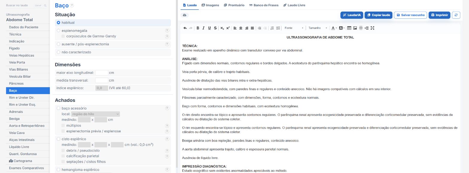
In the abdomen templates of Laudário, the spleen has its own tab, and there are templates for lymph nodes and for extended FAST (eFAST). In practice:
- Splenomegaly with longest axis, transverse measurement and splenic index.
- Accessory spleen as its own finding.
- Cysts, hemangiomas, abscesses, infarct and target lesions with ready-made phrases, including multiple lesions.
- Recommendations on a separate tab.
Try it for 15 days free, no credit card required.
The ultrasound reporting platform built by sonologists. We write about reports, classifications and standardization from the real routine of the practice. Who writes and how.
Educational content for healthcare professionals. It does not replace clinical judgment or the guidelines adopted by your service. Editorial policy.