Ultrasound for appendicitis: direct criteria, indirect findings and the appendix that was not seen
Diameter above 6 mm, compressibility, appendicolith and inflamed fat, signs of perforation and, above all, how to write the study in which the appendix was not characterized so that it is not read as negative.
 LaudárioUltrasound reports in practice
This article is part of the guide Abdominal Ultrasound: A Guide to the Findings That Actually Change Management.
Acute appendicitis is the most common abdominal surgical emergency, and ultrasound is the first imaging study in children, pregnant patients and thin young adults, precisely because it uses no radiation. But it is also one of the studies most dependent on the examiner and on the report: a "nonvisualized appendix" written ambiguously can be read as "appendicitis ruled out", and that is an error with consequences.
This text brings together the direct and indirect criteria, the technique that increases the visualization rate and the way to write each scenario, including the appendix that was not found.
The technique: graded compression
The technique described by Puylaert remains the basis of the study: with a high-frequency linear transducer, slow and progressive pressure is applied over the right iliac fossa, displacing gas-filled bowel loops until the psoas muscle and iliac vessels, which serve as landmarks, are exposed. The normal appendix is compressible; the inflamed one is not.
A few measures increase the chance of finding it:
- Ask the patient to point to the spot of greatest pain and start there.
- Search from the cecum, following the terminal ileum to the base of the appendix.
- Remember the atypical positions: retrocecal (left lateral decubitus helps), pelvic and subhepatic.
- In pregnant patients, the appendix rises with the uterus from the second trimester.
Direct criteria of appendicitis
- Transverse diameter greater than 6 mm, measured from serosa to serosa, with compression.
- Noncompressible structure, blind-ending, arising from the cecum.
- Thickened wall, with loss of layer stratification in more advanced cases.
- Mural hyperemia on Doppler, which may disappear in necrosis.
- Appendicolith: hyperechoic focus with acoustic shadowing in the lumen, which increases the risk of perforation and usually weighs on the surgical indication.
Diameter alone has a gray zone: between 6 and 7 mm, without other findings, interpretation requires caution, because normal appendices can reach this measurement. This is why indirect findings matter.
Indirect findings
- Hyperechoic, noncompressible periappendiceal fat: one of the strongest signs, because it shows inflammation extending around the appendix.
- Free fluid in the right iliac fossa or pelvis.
- Enlarged mesenteric lymph nodes in the right iliac fossa.
- Thickening of the cecum and terminal ileum.
- Tenderness on compression located exactly over the appendix (sonographic McBurney sign).
Signs of complication
- Perforation: wall discontinuity, loss of the appendiceal contour, periappendiceal collection, extraluminal appendicolith.
- Abscess: organized collection, with measurements and estimated volume. It changes management and may lead to drainage before surgery.
- Phlegmon: inflammatory mass involving bowel loops, omentum and appendix.
These findings should appear clearly in the diagnostic impression, because they change the type of approach.
The appendix that was not seen
This is the most delicate point of the report. The visualization rate varies widely with body habitus, appendix position and examiner experience. Three different scenarios need different wording:
- Appendix visualized, normal (compressible, up to 6 mm, no inflammatory signs): the study is reassuring.
- Appendix not visualized, no indirect findings: the study is inconclusive. It does not rule out appendicitis, and the report needs to say so. A sentence such as "appendix not characterized; no indirect findings of an inflammatory process in the right iliac fossa; the method does not exclude appendicitis" is honest and useful.
- Appendix not visualized, with indirect findings (inflamed fat, fluid, lymph nodes): should be described as findings consistent with an inflammatory process, with a recommendation for clinical correlation and, depending on the case, CT (or MRI in pregnant patients).
The error is writing "no signs of appendicitis" when the appendix was not seen.
Differential diagnoses the study can show
- Mesenteric adenitis, especially in children: enlarged lymph nodes clustered in the right iliac fossa, with a normal appendix.
- Terminal ileitis (infectious or Crohn disease).
- Right-sided colonic diverticulitis.
- Epiploic appendagitis: hyperechoic fatty nodule with a halo, tender on compression.
- In women: adnexal torsion, ruptured ovarian cyst, ectopic pregnancy. This is why pelvic assessment should be part of the study.
- Right ureteral lithiasis.
How to structure the report
- Technique (graded compression, linear transducer) and limitations (gas, pain, body habitus).
- Appendix: visualized or not; if visualized, diameter, compressibility, wall, Doppler and appendicolith.
- Indirect findings, one by one.
- Complications, with collection measurements.
- Diagnostic impression that answers the question: findings consistent with appendicitis, study without abnormalities or inconclusive study.
How Laudário helps
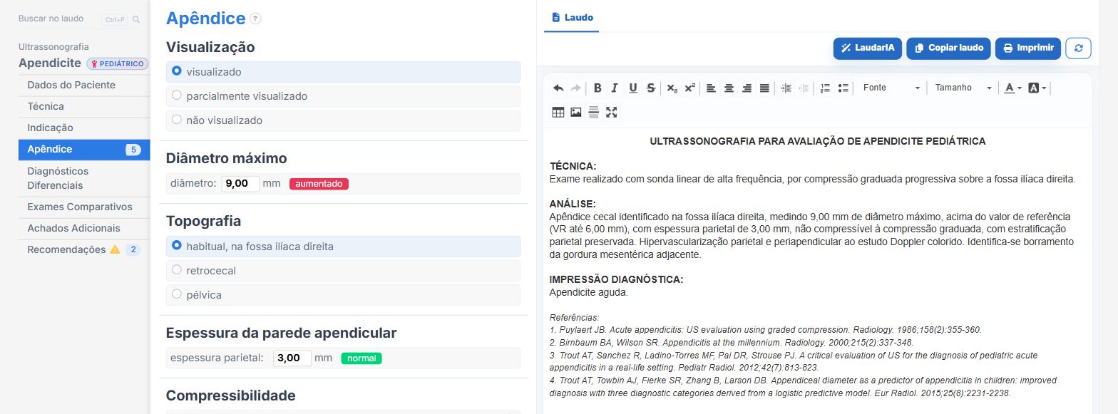
Laudário has a dedicated pediatric appendicitis template and abdomen templates, with fields for the right iliac fossa. In practice:
- Diameter, compressibility, wall, Doppler and appendicolith in their own fields.
- Indirect findings listed for checking, without relying on memory on call.
- Appendix visualization recorded, with the ready-made caveat that nonvisualization does not exclude acute appendicitis, so that an inconclusive study never goes out as negative.
Try it free for 15 days, no credit card required.
The ultrasound reporting platform built by sonologists. We write about reports, classifications and standardization from the real routine of the practice. Who writes and how.
Educational content for healthcare professionals. It does not replace clinical judgment or the guidelines adopted by your service. Editorial policy.