Adenomyosis on ultrasound: MUSA criteria, extent and the differential with fibroids
Direct and indirect signs by the MUSA consensus, diffuse or focal type, layer involved and extent, and the four elements that separate adenomyosis from a fibroid, including translesional vascularity.
 LaudárioUltrasound reports in practice
This article is part of the guide Gynecologic ultrasound: the complete guide, from uterus to ovaries.
For a long time adenomyosis was a diagnosis made on the surgical specimen: hysterectomy confirmed what the clinical picture suggested. Today transvaginal ultrasound makes this diagnosis with good accuracy, provided the examiner looks for the right signs and uses standardized language. It is also a frequent and underdiagnosed cause of heavy bleeding, severe cramping and infertility, often associated with endometriosis.
This text presents the criteria of the MUSA group, how to describe the extent of the disease, and the main differential diagnosis: the fibroid.
What it is and why ultrasound shows it
Adenomyosis is the presence of endometrial glands and stroma within the myometrium, with hypertrophy of the surrounding muscle. The sonographic signs reflect exactly this: islands of endometrium (echogenic or with small cysts) and reactive myometrium (thick, asymmetric, with shadowing).
The MUSA criteria
The MUSA consensus (Morphological Uterus Sonographic Assessment) organized the findings into direct signs, which show endometrial tissue in the myometrium, and indirect signs, which show the reaction of the myometrium.
Direct signs
- Myometrial cysts: small anechoic areas or areas with low-echogenicity content in the myometrium.
- Hyperechoic islands in the myometrium.
- Echogenic subendometrial lines and buds, which interrupt the junctional zone.
Indirect signs
- Wall asymmetry: one wall clearly thicker than the other, without a fibroid to explain it.
- Fan-shaped shadowing (hypoechoic linear striations).
- Globular, enlarged uterus.
- Translesional vascularity: on Doppler, the vessels cross the lesion perpendicular to the cavity, instead of curving around it as in a fibroid.
- Irregular or interrupted junctional zone, best assessed on three-dimensional ultrasound.
The direct signs are more specific. The update of the MUSA consensus proposes considering adenomyosis when there is at least one direct sign, and describing the indirect signs as suggestive when isolated. In practice, the more signs, the greater the confidence, and the report should state which were observed.
How to describe the extent
Saying only "signs of adenomyosis" does not allow comparison between studies or guide treatment. The consensus suggests describing:
- Location: anterior, posterior, fundal, lateral wall.
- Type: diffuse (spread through the myometrium) or focal (concentrated in one area); adenomyoma is the focal form surrounded by hypertrophic myometrium, with a nodular appearance.
- Layer involved: internal (junctional zone), middle or external. The external form is associated with deep endometriosis.
- Extent: proportion of the uterus involved, as mild, moderate or severe.
- Size of the focal lesions.
Adenomyosis or fibroid?
It is the most common differential and the one that most changes management, because fibroids can be resected and diffuse adenomyosis cannot be "enucleated".
- Margins: the fibroid is well defined; adenomyosis has ill-defined borders.
- Vascularity: circumferential, at the periphery, in the fibroid; translesional in adenomyosis.
- Mass effect: the fibroid displaces the endometrium and the serosa; adenomyosis barely deforms the uterine contour, despite enlarging the wall.
- Cysts and echogenic islands: typical of adenomyosis, rare in the fibroid.
The two conditions frequently coexist, and the report can and should describe both when present.
Adenomyosis and endometriosis
The association is frequent, especially external-layer adenomyosis with deep endometriosis of the posterior compartment. In patients with pelvic pain or infertility, an adenomyosis report should be accompanied by evaluation of the compartments. See our text on deep endometriosis mapping.
Common errors
- Diagnosing adenomyosis only from an enlarged uterus.
- Not differentiating the diffuse and focal types.
- Calling an adenomyoma a fibroid (or the reverse) without using Doppler.
- Not stating which signs were observed.
- Not evaluating associated endometriosis in a patient with pain.
How Laudário helps
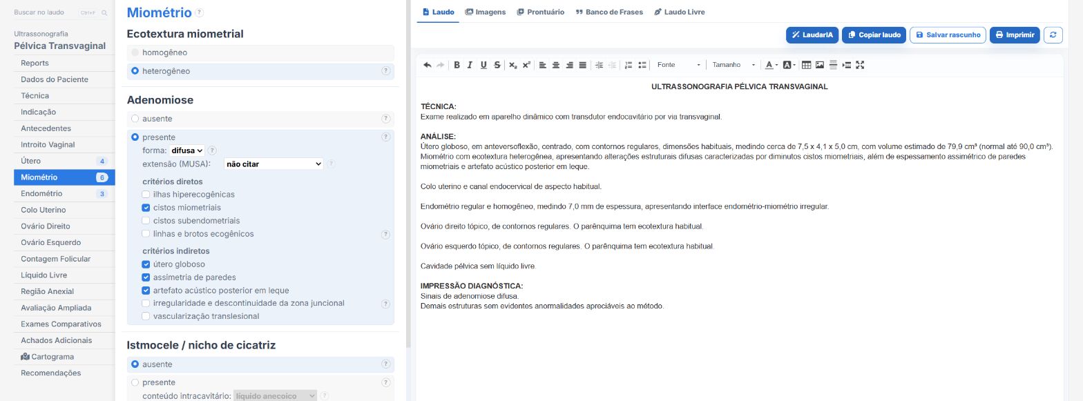
In the female pelvis and endometriosis templates of Laudário:
- Direct and indirect MUSA signs as options to check.
- Diffuse or focal type, location and extent.
- Fibroids in separate fields, so both findings coexist in the report.
- Recommendations in their own tab.
Try it for 15 days free, no credit card.
The ultrasound reporting platform built by sonologists. We write about reports, classifications and standardization from the real routine of the practice. Who writes and how.
Educational content for healthcare professionals. It does not replace clinical judgment or the guidelines adopted by your service. Editorial policy.