FIGO classification of fibroids: types 0 to 8 on ultrasound, mapping, and reporting
The FIGO classification of fibroids type by type, from 0 to 8: how many there are, where they are, and how much each invades the cavity. How to number and map when there are many, and how to differentiate from adenomyosis, polyp, and adnexal mass.
 LaudárioUltrasound reports in practice
This article is part of the guide Gynecologic ultrasound: the complete guide, from uterus to ovaries.
Uterine fibroids are the most common pelvic finding in adult women, and also one of the most poorly described. "Fibroid uterus" or "multiple myometrial nodules" does not tell the gynecologist what is needed to decide between observation, medical treatment, hysteroscopy, myomectomy, or embolization. That decision depends on three pieces of information that ultrasound delivers well: how many, where, and how much each one invades the cavity.
The FIGO classification answers the third question: it numbers the fibroid from 0 to 8 according to its position relative to the cavity and the serosa. Types 0 to 2 are submucosal, 3 to 4 are intramural, and 5 to 7 are subserosal; when a fibroid touches both the cavity and the serosa, two numbers are used, such as FIGO 2-5. Below are the types one by one, followed by how to describe, measure, and map fibroids in the report.
FIGO classification of fibroids: types 0 to 8
The FIGO classification describes the position of the fibroid relative to the cavity and the serosa, in types 0 to 8:
- Type 0: pedunculated submucosal, entirely within the cavity.
- Type 1: submucosal, with less than 50% in the myometrium.
- Type 2: submucosal, with 50% or more in the myometrium.
- Type 3: intramural, in contact with the endometrium, without deforming the cavity.
- Type 4: purely intramural.
- Type 5: subserosal, with 50% or more intramural.
- Type 6: subserosal, with less than 50% intramural.
- Type 7: pedunculated subserosal.
- Type 8: other (cervical, parasitic, broad ligament).
Fibroids that touch both the cavity and the serosa are described with two numbers, such as 2-5. The practical importance is direct: types 0, 1, and 2 are candidates for hysteroscopy and are related to bleeding and infertility; subserosal fibroids rarely cause bleeding, but can cause compression.
The typical appearance
The leiomyoma appears as a solid, well-defined nodule, usually hypoechoic or heterogeneous, with fan-shaped acoustic shadowing and peripheral, circumferential vascularity on Doppler. These elements form the typical appearance described by the MUSA group (Morphological Uterus Sonographic Assessment), which standardized myometrial terminology.
Fibroids can calcify (hyperechoic image with dense shadowing, common after menopause) or undergo degeneration, with cystic areas and heterogeneity. Degeneration, especially in pregnancy, explains acute pain.
What to measure and record for each fibroid
- Location: wall (anterior, posterior, fundal, right or left lateral) and segment (corpus, isthmus, cervix).
- FIGO type.
- Measurements in all three axes.
- For submucosal fibroids, the thickness of free myometrium between the fibroid and the serosa, which informs the safety of hysteroscopy.
- Calcification, degeneration, and vascularity, when relevant.
In addition to the fibroids, the report records the total uterine volume and the endometrium, which may be deformed or difficult to measure.
Uterus with many fibroids: mapping
With several nodules, describing all of them in running text produces an unreadable paragraph. The solution is to number the fibroids (F1, F2, F3...) and keep the same numbering in follow-up exams, presenting each one on its own line or in a table. When there are many, an accepted alternative is to describe individually the largest ones and those related to the cavity, and group the small intramural ones ("in addition to multiple smaller intramural nodules under 1 cm").
A map (cartogram) of the uterus with the position of each fibroid is what the surgeon values most in planning a myomectomy.
Differential diagnoses
- Adenomyosis: poorly defined lesion, without a capsule, with vessels crossing the lesion (rather than encircling it), myometrial cysts, and striations. Focal adenomyoma is the most difficult differential.
- Endometrial polyp: differentiated from a type 0 fibroid by its single feeding vessel and by echogenicity.
- Solid adnexal mass: a pedunculated subserosal fibroid can mimic an ovarian tumor. Identifying the ovary as separate from the lesion and the vascular bridge to the uterus resolves it.
- Leiomyosarcoma: rare, but should be considered in the face of rapid growth after menopause, a large heterogeneous lesion with irregular cystic areas and disorganized central vascularity. In these situations, the recommendation for MRI is part of the report.
Common errors
- "Fibroid uterus" without number, location, and type.
- Submucosal fibroid without the FIGO type and without the free myometrium.
- Different numbering between exams, which prevents comparing growth.
- Pedunculated fibroid mistaken for an adnexal mass.
- Not mentioning the relationship with the cavity in a patient with bleeding or infertility.
For the complete structure of the exam, see our text on the transvaginal pelvic report.
How Laudário helps
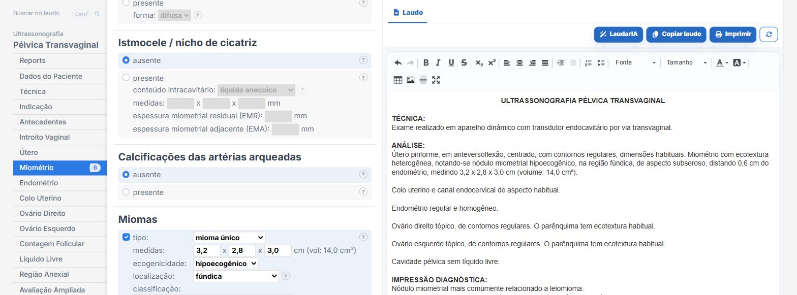
In the female pelvis templates of Laudário, fibroids are added one by one. In practice:
- Each fibroid with numbering, location, measurements, and FIGO type.
- The same fields for follow-up, keeping the numbering.
- Calculated uterine volume.
- Adenomyosis by MUSA criteria in its own fields, so the differential is clear.
Try it for 15 days free, no credit card required.
The ultrasound reporting platform built by sonologists. We write about reports, classifications and standardization from the real routine of the practice. Who writes and how.
Educational content for healthcare professionals. It does not replace clinical judgment or the guidelines adopted by your service. Editorial policy.